article thumbnail

Can We Upskill Radiographers through Artificial Intelligence? 

The British Institute of Radiology

Shamie Kumar describes how AI fits into a radiology clinical workflow and her perspective on how a clinical radiographer could use this to learn from and enhance their skills. If the AI findings are seen in PACS, how many radiographers actually log into PACS after taking a scan or X-ray? Can Radiographers Up-Skill?

article thumbnail

Artificial Intelligence Embedded Imaging Modality

The British Institute of Radiology

In the third blog of her series on AI and the radiographer, Shamie Kumar explores the impact on the radiographer when AI is integrated within an imaging modality. The question to explore in this blog is when AI is integrated within an imaging modality itself and how that may impact a radiographer.

Insiders

Sign Up for our Newsletter

This site is protected by reCAPTCHA and the Google Privacy Policy and Terms of Service apply.

Trending Sources

article thumbnail

Is Artificial Intelligence a glorified red dot system?

The British Institute of Radiology

But how will AI in the workplace affect the radiographer and how does it differ from the red dot system radiographers are so familiar with? The Red Dot System Often one of the first courses a newly qualified radiographer attends is the red dot course. What does AI do that a radiographer doesn’t already?

article thumbnail

WBCT Indication Series: Hallux Valgus

CurveBeam AI

Diagnosis While radiographs are typically sufficient to make the diagnosis, WBCT scans may be useful to plan surgical treatment. Accurately assess sesamoid position as plain radiographs cannot determine whether the sesamoids have been reduced within their grooves 5. JBJS 2009. (3) Conti et al. FAI 2022. (2) Okuda et al.

article thumbnail

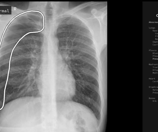
The Needle Vs. The Knife for Spontaneous Pneumothorax: A Closer Look at the EXPRED Study

REBEL EM

Sites: Investigators recruited patients at 31 French emergency departments at university and nonuniversity hospitals Duration : June 1, 2009 to March 31, 2015. Absence of residual pneumothorax or the presence of an apical residual pneumothorax smaller than 2 cm, based on chest radiography. Recurrence of pneumothorax within 1 year.