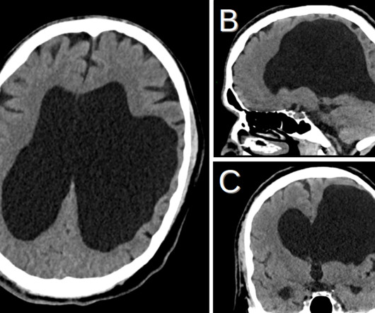
Porencephaly
Global Radiology CME
JULY 26, 2021
On MRI, the cystic components will appear as low signal intensity on T1 weighted images, high signal intensity on T2 weighted images, low signal intensity on FLAIR images, and will have no restricted diffusion on DWI [10-12]. Reproductive and Developmental Toxicology, Elsevier, 2011, pp. Contro, Elena, et al.


Let's personalize your content