Remove 2011 Remove Radiograph Remove Radiographer
article thumbnail

Weight Bearing CT for Foot & Ankle Imaging

CurveBeam AI

Common Indications Syndesmosis Provide increased sensitivity and specificity over radiographs 1. Flat Foot Provide an assessment of important anatomical markers of pronounced hindfoot deformity and peritalar subluxation (PTS), difficult to visualize on conventional two-dimensional radiographs 2. 2011 Feb;28(2):195-8. Diabet Med.

article thumbnail

WBCT Indication Series: Charcot Arthropathy

CurveBeam AI

Non-WB radiographs could not determine instability or severity of midfoot collapse; surgical intervention postponed. One year later, deformity had significantly progressed, but the remaining bone stock was unclear from plain radiographs.​ WB radiographs of the left foot demonstrated Charcot arthropathy. 2011 Feb;28(2):195-8.

Insiders

Sign Up for our Newsletter

This site is protected by reCAPTCHA and the Google Privacy Policy and Terms of Service apply.

Trending Sources

article thumbnail

Plastic Bowing Fracture Ulna

Global Radiology CME

Figure 2 A: AP view radiograph of right forearm. B: Lateral radiograph view of right forearm. An angulated fracture of the distal midshaft radius is also visualized, but there is also bowing of the ulna that is more appreciated on the lateral radiograph view. 8 Year Old Male With Trauma Due To A Fall. Xray of the Week Figure 1.

article thumbnail

AI model shows promise for interpreting follow-up chest x-rays

AuntMinnie

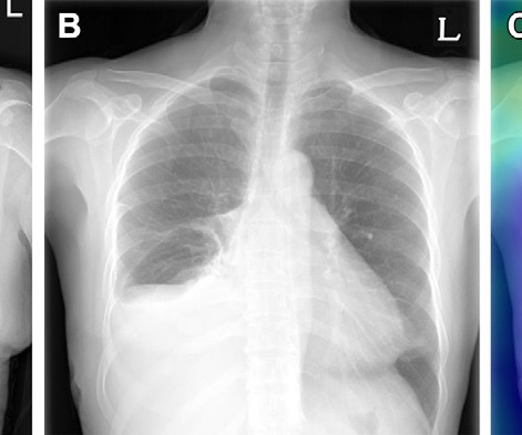
Radiologists routinely compare the current and previous chest radiographs during interpretation to enhance the sensitivity for change detection and provide information for differential diagnosis. Example of triage of no change in a pair of chest radiographs in the emergency department. (A) Image courtesy of Radiology.

X-ray 111
article thumbnail

Teleradiology Proves as a Reliable, Efficient and Economically Viable Services for All Stakeholders

Cloudex Radiology

Our team stands up to the statement on quality as briefed in Radiographics, 2011.

article thumbnail

Porencephaly

Global Radiology CME

Reproductive and Developmental Toxicology, Elsevier, 2011, pp. Seeing the radiographic images made medical education come to life for him. Avery’s Diseases of the Newborn, Elsevier, 2018, pp. doi: 10.1016/B978-0-323-40139-5.00059-0. Mikic, Aleksandra Novakov, et al. doi: 10.1016/B978-0-12-382032-7.10074-8. Contro, Elena, et al.

article thumbnail

A brief history of radiology

Radiology Cafe

Photoprint from radiograph by W.K. 3) In the early twentieth century, it was a common goal for investigators to try to find a way to separate the superimposed shadows that were recorded when a complex structure was shown on a radiograph. (3) “First MRI and ultrasound scanning” Archived from the original on 2011-11-20.