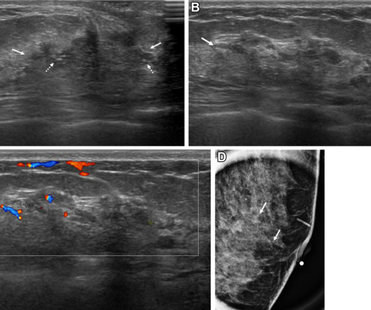
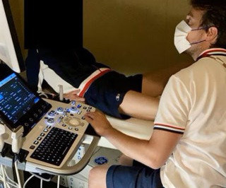
article thumbnail

Diagnostic Imaging Trends: Point of Care Ultrasound – POCUS

Vesta Teleradiology

Point of care ultrasound (POCUS) is revolutionizing the healthcare industry and changing how doctors prescribe treatment for patients. POCUS is a diagnostic tool that utilizes ultrasound imaging to diagnose, monitor, and guide treatments for medical conditions. What Is Point of Care Ultrasound (POCUS)?

article thumbnail

Lymphoscintigraphy underused in the U.S. to diagnose lymphedema

AuntMinnie

Several recent expert panels have suggested from anecdotal experience that LSG is used infrequently and that the diagnosis of lymphedema is usually based on clinical examination, the team wrote. insurance database who had a new diagnosis of lymphedema between April 2012 to March 2020.

Diagnose 119
Insiders

Sign Up for our Newsletter

This site is protected by reCAPTCHA and the Google Privacy Policy and Terms of Service apply.

Trending Sources

article thumbnail

Olympic imaging squad gets set for summer games

AuntMinnie

Clinical information is essential to refine the diagnosis, but we must also understand the sporting issues in order to adapt the athletes’ care.” Renoux has accompanied the French team to the Olympics since London 2012, and since 2006, he has also been part of the radiology team covering the French Open tennis tournament at Roland Garros.

Imaging 75
article thumbnail

REBEL Core Cast 116.0 – Achilles Tendon Rupture

REBEL EM

Take Home Points Achilles tendon rupture is a clinical diagnosis. You will notice the squeeze will cause no motion if there is a full rupture/tear, and diminished motion if there is a partial tear Performance Characteristics ( Garras 2012 ) Sensitivity Specificity (+) LR (-) LR 96-100% 93-100% 13.7 Br J Sports Med. Coll Antropol.

X-ray 77
article thumbnail

REBEL Core Cast 85.0 – Superficial Venous Thrombosis

REBEL EM

Bauersachs 2013 ) Clinical Significance SVT previously thought to be a benign disease entity ( Cosmi 2015 ) However, subsequent studies have demonstrated that in patients with lower extremity SVT concomitant DVT and PE was seen in approximately 25% and 5% respectively. Risk factors for the development of SVT are similar to those of VTE.

Disease 52
article thumbnail

Don’t Forget About the IO in the Critically Ill Patient

REBEL EM

The current standard of practice has moved away from landmark-based central line placement given the efficacy and safety of ultrasound-based techniques. Clinical Management of Intraosseous Access in Adults in Critical Situations for Health Professionals. 2012 PMID: 21893125. minutes compared to a mean placement time of 10.7

Hospital 104
article thumbnail

REBEL Core Cast 97.0 – Acute Vision Loss II

REBEL EM

Signs Shafer’s sign (tobacco dust) May see a Weiss ring when the posterior vitreous (PV) detaches from the optic disc margin Visualization with ultrasound. et al, Emergency Medicine Clinical Essentials ed 2. et al, Emergency Medicine Clinical Essentials ed 2. Emergency Department Management: Emergency ophthalmology consultation.