This site uses cookies to improve your experience. To help us insure we adhere to various privacy regulations, please select your country/region of residence. If you do not select a country, we will assume you are from the United States. Select your Cookie Settings or view our Privacy Policy and Terms of Use.
Cookie Settings
Cookies and similar technologies are used on this website for proper function of the website, for tracking performance analytics and for marketing purposes. We and some of our third-party providers may use cookie data for various purposes. Please review the cookie settings below and choose your preference.
Used for the proper function of the website
Used for monitoring website traffic and interactions
Cookie Settings
Cookies and similar technologies are used on this website for proper function of the website, for tracking performance analytics and for marketing purposes. We and some of our third-party providers may use cookie data for various purposes. Please review the cookie settings below and choose your preference.
Strictly Necessary: Used for the proper function of the website
Performance/Analytics: Used for monitoring website traffic and interactions
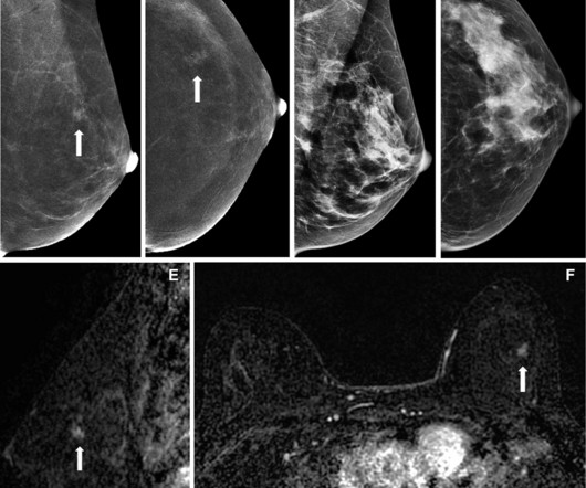
They compared the respective performances of CEM, low-energy mammograms alone, and low-energy imaging supplemented by whole-breast ultrasound. Their study included data collected between 2014 and 2019 from 468 women with a median age of 54. However, it also led to more biopsy recommendations. Abnormal interpretation rate 10.3%
Though the initial focus of the FAST exam was for detection of abdominal free fluid, the eFAST (Extended FAST) is more commonly used and adds thoracic windows helping to identify pneumothorax ( Musthafa 2014 ). All images were then uploaded for attending radiologists to read in real time.
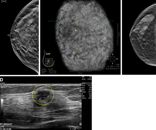
(D) Antiradial gray-scale image in the right breast from supplemental handheld screening ultrasound in the same 74-year-old patient 7 months later demonstrates an irregular hypoechoic mass in the right breast (yellow outline), which yielded a diagnosis of invasive ductal carcinoma. Image courtesy of the RSNA.
This means the radiologists have to be on their game all the time with an ever increasing workload compounded by complexity of available imagingmodalities such as 3D mammography. This is set against a global backdrop where there just aren’t enough breast radiologists available and burnout is on the rapid rise.
We organize all of the trending information in your field so you don't have to. Join 5,000 users and stay up to date on the latest articles your peers are reading.
You know about us, now we want to get to know you!
Let's personalize your content
Let's get even more personalized
We recognize your account from another site in our network, please click 'Send Email' below to continue with verifying your account and setting a password.


Let's personalize your content