Remove 2014 Remove Magnetic Resonance Imaging Remove Radiograph Remove Radiographer
article thumbnail

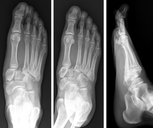
Lisfranc Fracture Dislocation

Global Radiology CME

A) AP radiograph of Lisfranc Fracture Dislocation demonstrates the circled “fleck sign” or Lisfranc ligament avulsion fracture fragment. (B) C) The lateral radiograph notes with a circle, the dorsal sub dislocation of the metatarsal base. Radiographs should be repeated after two weeks to ensure surgery is unnecessary.

article thumbnail

Journal Scan – This Month in Other Journals, June 2023

AJNR

It has well-defined radiographic features and various clinical presentations. They described six radiographic phenotypes of CSVD: (1) recent small subcortical infarct, (2) white matter hyperintensity, (3) lacune of presumed vascular origin, (4) widened perivascular spaces, (5) cerebral microbleed, and (6) brain atrophy.

Disease 40
article thumbnail

Emergency Guide to Stroke Neuroimaging

Northwestern EM

A 2014 pooled analysis of 21 studies from 1993 to 2013 showed CTA has a sensitivity of 83.2% 2014 Nov 29;384(9958):1929-35. Magnetic resonance imaging and computed tomography in emergency assessment of patients with suspected acute stroke: a prospective comparison. Quant Imaging Med Surg. Radiographics.