Remove 2014 Remove Radiograph Remove Radiographer Remove Radiography
article thumbnail

Opportunity for osteoporosis check on lumbar spine plain radiographs

AuntMinnie

. | W3-SSMK08-4 | Room E450A A deep learning-based framework for automated screening of osteoporosis on lumbar spine plain radiographs shows potential as another way to opportunistically make use of imaging studies performed for other indications, according to this presentation.

article thumbnail

Lisfranc Fracture Dislocation

Global Radiology CME

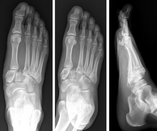
A) AP radiograph of Lisfranc Fracture Dislocation demonstrates the circled “fleck sign” or Lisfranc ligament avulsion fracture fragment. (B) C) The lateral radiograph notes with a circle, the dorsal sub dislocation of the metatarsal base. Radiographs should be repeated after two weeks to ensure surgery is unnecessary.

article thumbnail

The Needle Vs. The Knife for Spontaneous Pneumothorax: A Closer Look at the EXPRED Study

REBEL EM

Thoracentesis with free drainage for 15 minutes Drainage at -25cm H20 for 30 minutes Clamping and radiograph; If failed, 2nd aspiration at -25cm H20 for 30 minutes, Repeat clamping and radiograph. Absence of residual pneumothorax or the presence of an apical residual pneumothorax smaller than 2 cm, based on chest radiography.