This site uses cookies to improve your experience. To help us insure we adhere to various privacy regulations, please select your country/region of residence. If you do not select a country, we will assume you are from the United States. Select your Cookie Settings or view our Privacy Policy and Terms of Use.
Cookie Settings
Cookies and similar technologies are used on this website for proper function of the website, for tracking performance analytics and for marketing purposes. We and some of our third-party providers may use cookie data for various purposes. Please review the cookie settings below and choose your preference.
Used for the proper function of the website
Used for monitoring website traffic and interactions
Cookie Settings
Cookies and similar technologies are used on this website for proper function of the website, for tracking performance analytics and for marketing purposes. We and some of our third-party providers may use cookie data for various purposes. Please review the cookie settings below and choose your preference.
Strictly Necessary: Used for the proper function of the website
Performance/Analytics: Used for monitoring website traffic and interactions
Conversely, thermal ablation – radiofrequency ablation, microwave ablation, and laser ablation – are minimally invasive procedures guided by ultrasound that use extreme temperatures to destroy the tumors. Studies have shown favorable results of the procedures, yet to date, long-term outcomes have not been reported, the authors noted.
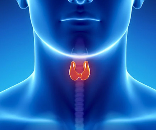
A nomogram combining ultrasound features and TI-RADS parameters can differentiate between malignant and benign thyroid nodules, according to research published January 22 in Ultrasound in Medicine & Biology. The American College of Radiology (ACR) created TI-RADS based on gray-scale ultrasound features.
Our top article in 2014 reported on the financial benefits of health information exchanges. The researchers retrospectively analyzed repeat imaging rates for CT, chest x-ray, and ultrasound in 37 emergency departments in California and Florida that have initiated HIE. of ultrasound cases), and 29,073 repeat chest x-rays (19.5%
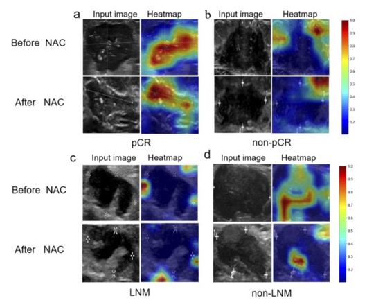
An ultrasound-based deep learning radiomics nomogram (DLRN) model performed well in a study about predicting tumor status and lymph node metastasis in breast cancer patients post chemotherapy. They added to the literature, exploring the feasibility of an ultrasound-based fusion model combining the aforementioned features.
An algorithm combining the Fibrosis-4 Index (FIB-4) and an ultrasound deep-learning model could improve diagnostic accuracy and referral management for all-cause advanced liver fibrosis, a study published April 23 in Radiology found. Chen and colleagues constructed sequential clinical algorithms that include an ultrasound deep-learning model.
A team led by Joao Horvat, MD, from the Memorial Sloan Kettering Cancer Center in New York found that CEM depicted 90% of breast cancers compared with 10% on low-energy mammograms alone and 50% on low-energymammogramswith whole-breast ultrasound. Their study included data collected between 2014 and 2019 from 468 women with a median age of 54.
Elliot Fishman, MD, Johns Hopkins Medicine The second finalist is Elliot Fishman, MD, a category for which he took the trophy in 2001, 2007, 2014, and 2017. Last year, he was awarded a grant through the American Roentgen Ray Society's (ARRS) clinician educator development program.
Background: Point-of-care ultrasound (PoCUS) is a valuable clinical tool in the assessment of acute dyspnea. Impact of serial cardiopulmonary point-of-care ultrasound exams in patients with acute dyspnoea: a randomized, controlled trial. PoCUS evaluations included lung ultrasound (LUS) and focused cardiac ultrasound (FoCUS).
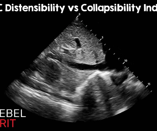
Regarding caval indexes, the advent of artificial intelligence and advanced learning has become integrated into many ultrasound machines. Ultrasound Med Biol. May 2014; PMID: 24495437. As with most things in medicine, it is important to understand the pitfalls. Most measurements are somewhere around the hepatic confluence.
Dr. David Gauden, a co-founder of Blue Earth Diagnostics and with the company since 2014, assumes the role of full-time CEO of Bracco subsidiary Blue Earth Therapeutics. Board of Directors and will serve as Vice Chair of the Blue Earth Diagnostics Inc. Board of Directors. Bracco Imaging S.p.A.,
The 2014 CPG for management of VLUs recommends performing a venous reflux duplex ultrasound (VRDUS) and prescribing pentoxifylline. Studies have identified several barriers that can reduce primary care clinician adherence to clinical practice guidelines (CPGs).
Background: The use of ultrasound is well established for trauma patients in the emergency department, with almost every patient receiving a FAST (Focused Assessment with Sonography in Trauma) examination as part of the “ABC’s” of trauma. Not so FAST- Chest ultrasound underdiagnoses traumatic pneumothorax. PMID: 34932040.
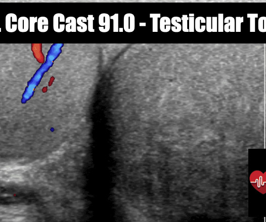
Ultrasound of acute vasitis shows a heterogenous and thickened vas deferens. Color Doppler ultrasound of the same section as Figure 4. With ultrasound, acute vasitis shows a heterogenous, hypoechoic and thickened vas deferens with hyperemia on color Doppler (3-6)(Figs. Journal of Ultrasound in Medicine. J Ultrasound Med.
History, physical examination and ultrasound are all flawed in making the diagnosis. History, physical examination and ultrasound are all flawed in making the diagnosis. 2014, (Ch) 99: p 1326-1356. The gold standard is surgical exploration. Physical exam, history and imaging all have significant limitations. 10, (-) LR = 0.13
Frappe 2014, Cosmi 2015 ) Half of the DVTs seen in SVT are non-contiguous and 17% of DVTs are seen in the contralateral limb. If an SVT is uncovered in the lower extremity, a bilateral duplex ultrasound evaluating the deep venous system should be considered. J Thromb Haemost 2014; 12: 831–8. PMID: 24679145 White RH.
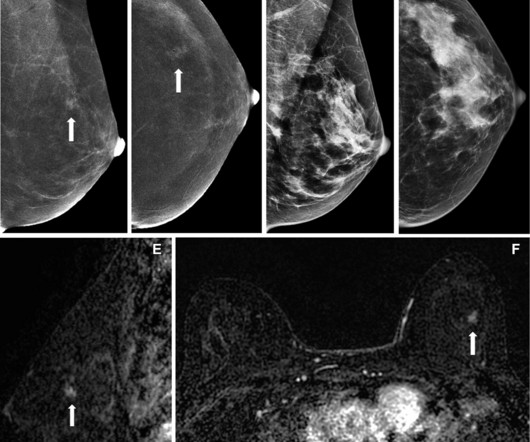
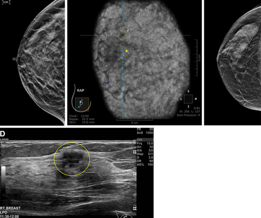
Supplemental breast ultrasound may have utility in imaging women with dense breasts and high risk of advanced or invasive breast cancer, a study published August 6 in Radiology found. Sample images show cancer detection at supplemental ultrasound screening after screening mammography with a negative result. (A)
While her mammogram yielded negative results, a subsequent ultrasound found breast cancer. Two barriers include access to follow-up imaging such as MRI and ultrasound, and insurance coverage for supplemental imaging. While September 10 may be a day to celebrate, breast imaging advocates say there is much work ahead.
Randomized, Controlled Trial of Ultrasound-Assisted Catheter-Directed Thrombolysis for Acute Intermediate-Risk Pulmonary Embolism. A prospective, Single-Arm Multicenter Trial of Ultrasound-Facilitated, Catheter-Directed, Low-Dose Fibrinolysis for Acute Massive and Submassive Pulmonary Embolism: The SEATTLE II Study. CHEST 2010.
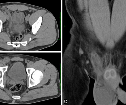
Ultrasound of gallbladder used for guidance of percutaneous needle (red arrow) placement for cholecystostomy. Ultrasound of gallbladder demonstrating drainage catheter in the lumen (blue arrow). Technique and indications of percutaneous cholecystostomy in the management of acute cholecystitis in 2014. Hojberg and Caliskan].
Philadelphia, PA: Elsevier Saunders; 2014: 2282-2299. Gorgas, D. “Infections related to pregnancy.” ” Emerg Med Clin North Am 26 (2): 345-366, viii. PMID: 18406978 Houry, D and B. Acute Complications of Pregnancy. In: Marx, J et al, ed. Rosen’s Emergency Medicine. Post Peer Reviewed By: Salim R.
We organize all of the trending information in your field so you don't have to. Join 5,000 users and stay up to date on the latest articles your peers are reading.
You know about us, now we want to get to know you!
Let's personalize your content
Let's get even more personalized
We recognize your account from another site in our network, please click 'Send Email' below to continue with verifying your account and setting a password.


Let's personalize your content