Remove 2015 Remove Clinic Remove Computed Tomography Remove Radiographer
article thumbnail

Lisfranc Fracture Dislocation

Global Radiology CME

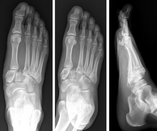
A) AP radiograph of Lisfranc Fracture Dislocation demonstrates the circled “fleck sign” or Lisfranc ligament avulsion fracture fragment. (B) C) The lateral radiograph notes with a circle, the dorsal sub dislocation of the metatarsal base. Radiographs should be repeated after two weeks to ensure surgery is unnecessary.

article thumbnail

WBCT Indication Series: Hallux Valgus

CurveBeam AI

Diagnosis While radiographs are typically sufficient to make the diagnosis, WBCT scans may be useful to plan surgical treatment. Accurately assess sesamoid position as plain radiographs cannot determine whether the sesamoids have been reduced within their grooves 5. . • Assess congruency and degenerative changes at the 1st MTP joint.

Insiders

Sign Up for our Newsletter

This site is protected by reCAPTCHA and the Google Privacy Policy and Terms of Service apply.

Trending Sources

article thumbnail

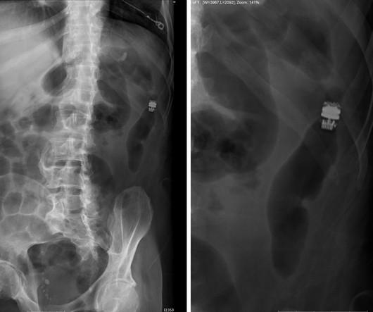
Capsule Retention Following Capsule Endoscopy

Global Radiology CME

Frontal abdomen radiograph demonstrates foreign body consistent with capsule endoscopy device (pill cam) in descending colon. Approximately 2% of all capsule endoscopies result in CR [ 2 ] The clinical indication for capsule endoscopy is correlated with different rates of CR. 2015.3820/2015 9. Xray of the Week Figure 1.

X-ray 52
article thumbnail

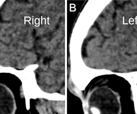
Lens Subluxation

Global Radiology CME

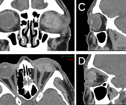
Computed tomography is also an alternative method for lens subluxation which again can show deviation of the lens (Figs. Radiographics. Sai is also a member of the Gold Humanism Honor Society and is involved with giving back to the community at a local free clinic as a medical assistant. 2006;10(4):345-350.

article thumbnail

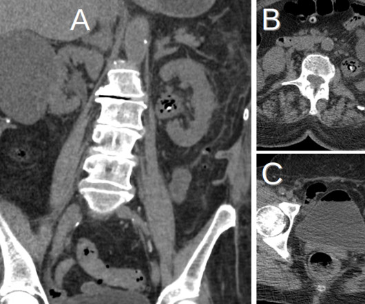
Emphysematous Pyelonephritis

Global Radiology CME

KUB indicates kidneys, ureter, and bladder (plain abdominal radiograph); CT, computed tomography; and PCD, percutaneous catheter drainage. In severe cases or patients who do not respond to PCD, treatment with nephrectomy can lead to clinical and radiological improvement (Fig. RadioGraphics. 2000;160(6):797-805.

article thumbnail

A brief history of radiology

Radiology Cafe

Photoprint from radiograph by W.K. 3) In the early twentieth century, it was a common goal for investigators to try to find a way to separate the superimposed shadows that were recorded when a complex structure was shown on a radiograph. (3) 3) This is what is known as tomography. This is now known as ‘Hand mit Ringen’. (1)

article thumbnail

Ruptured Globe

Global Radiology CME

1 Imaging: Computed tomography (CT) is the recommended imaging modality for evaluating orbital trauma. Clinical features of single and repeated globe rupture after penetrating keratoplasty. Radiographics. Penetrating and blunt trauma, in addition to chemical exposure, account for the majority of cases. Clin Ophthalmol.