Remove 2015 Remove Computed Tomography Remove Magnetic Resonance Imaging
article thumbnail

Lisfranc Fracture Dislocation

Global Radiology CME

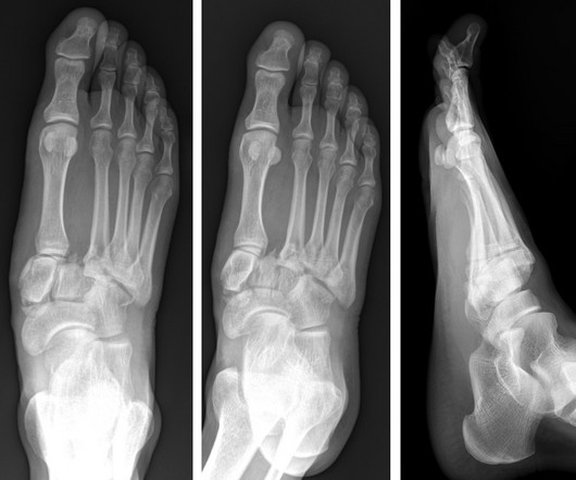
A computed tomography scan will better assist with diagnosis and help with planning if surgery is necessary [3]. Magnetic resonance imaging will help to evaluate ligamentous injury and provides a 94% predictive value for diagnosing Lisfranc injury [5]. J Foot Ankle Res. 2023;16(1):9. Published 2023 Mar 1.

article thumbnail

Type 3 Cuboid Fracture

Global Radiology CME

More sophisticated imaging, such as computed tomography or magnetic resonance imaging, should be obtained if plain film is unrevealing but there is high suspicion of fracture. Diagram showing the types of fracture of the cuboid [1].

Insiders

Sign Up for our Newsletter

This site is protected by reCAPTCHA and the Google Privacy Policy and Terms of Service apply.

Trending Sources

article thumbnail

A brief history of radiology

Radiology Cafe

Tomography originates from the Greek words ‘tomos’, meaning ‘slice’ or ‘section’, and ‘graphia’, meaning ‘description of’. In the 1960s computers were increasingly available and more powerful and in 1971, the first computed tomography (CT) scan was performed on a patient. Tumor detection by nuclear magnetic resonance.

article thumbnail

The radiologist in 22 years time

Radiology Cafe

A trial, which involved analysing a computerised tomography (CT) scan of a patient’s lungs against three expert human radiologists, found that Enlitic’s system was 50% better at classifying malignant tumours and had a false-negative rate of zero, compared with 7% for the humans (3).

article thumbnail

Journal Scan – This Month in Other Journals, June 2023

AJNR

Patients had a large ischemic-core volume, defined as an ASPECT score of 3 to 5 (range, 0 to 10, with lower scores indicating larger infarction) or a core volume of at least 50 ml on computed tomography perfusion or diffusion-weighted magnetic resonance imaging. 3 tables, 1 figure, no images 6.

Disease 40
article thumbnail

Emergency Guide to Stroke Neuroimaging

Northwestern EM

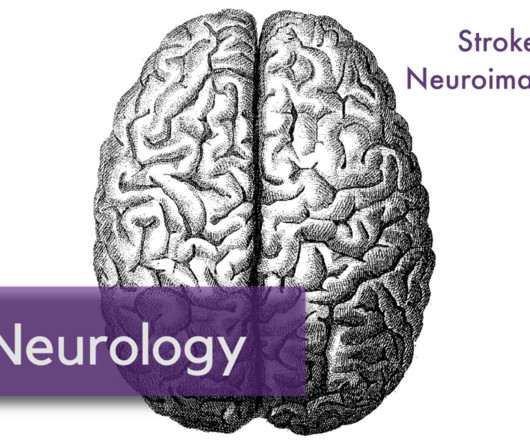
As these two time-windows overlap, the most practical approach is increasingly to obtain multi-modality imaging up-front / as rapidly as possible in the ED. Magnetic resonance imaging and computed tomography in emergency assessment of patients with suspected acute stroke: a prospective comparison.