Remove 2015 Remove Computed Tomography Remove Magnetic Resonance Imaging Remove Radiographer
article thumbnail

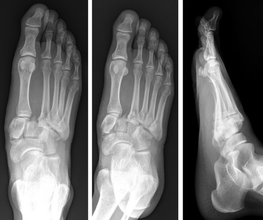
Lisfranc Fracture Dislocation

Global Radiology CME

A) AP radiograph of Lisfranc Fracture Dislocation demonstrates the circled “fleck sign” or Lisfranc ligament avulsion fracture fragment. (B) C) The lateral radiograph notes with a circle, the dorsal sub dislocation of the metatarsal base. Radiographs should be repeated after two weeks to ensure surgery is unnecessary.

article thumbnail

Type 3 Cuboid Fracture

Global Radiology CME

A) Dorsoplantar radiograph of the foot demonstrating an isolated fracture of the cuboid with possible extension into the tarsometatarsal joint. (B) B) Medial oblique radiograph of the foot demonstrating an isolated fracture of the cuboid. Radiographic evidence can support the diagnosis. Trauma in a 8 year old female.

Insiders

Sign Up for our Newsletter

This site is protected by reCAPTCHA and the Google Privacy Policy and Terms of Service apply.

Trending Sources

article thumbnail

A brief history of radiology

Radiology Cafe

after seeing the image. (2) Photoprint from radiograph by W.K. 3) In the early twentieth century, it was a common goal for investigators to try to find a way to separate the superimposed shadows that were recorded when a complex structure was shown on a radiograph. (3) 3) This is what is known as tomography. Alan Chodos.

article thumbnail

The radiologist in 22 years time

Radiology Cafe

A trial, which involved analysing a computerised tomography (CT) scan of a patient’s lungs against three expert human radiologists, found that Enlitic’s system was 50% better at classifying malignant tumours and had a false-negative rate of zero, compared with 7% for the humans (3). 2011, Radiographics, pp. Hough, Andrew.

article thumbnail

Journal Scan – This Month in Other Journals, June 2023

AJNR

It has well-defined radiographic features and various clinical presentations. They described six radiographic phenotypes of CSVD: (1) recent small subcortical infarct, (2) white matter hyperintensity, (3) lacune of presumed vascular origin, (4) widened perivascular spaces, (5) cerebral microbleed, and (6) brain atrophy.

Disease 40
article thumbnail

Emergency Guide to Stroke Neuroimaging

Northwestern EM

As these two time-windows overlap, the most practical approach is increasingly to obtain multi-modality imaging up-front / as rapidly as possible in the ED. Magnetic resonance imaging and computed tomography in emergency assessment of patients with suspected acute stroke: a prospective comparison.