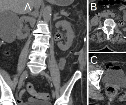
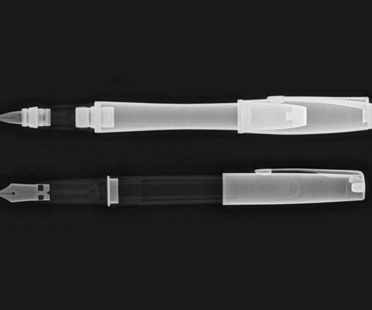
Remove 2015 Remove Computed Tomography Remove Radiograph
article thumbnail

WBCT Indication Series: Hallux Valgus

CurveBeam AI

Diagnosis While radiographs are typically sufficient to make the diagnosis, WBCT scans may be useful to plan surgical treatment. Accurately assess sesamoid position as plain radiographs cannot determine whether the sesamoids have been reduced within their grooves 5. . • Assess congruency and degenerative changes at the 1st MTP joint.

article thumbnail

Lisfranc Fracture Dislocation

Global Radiology CME

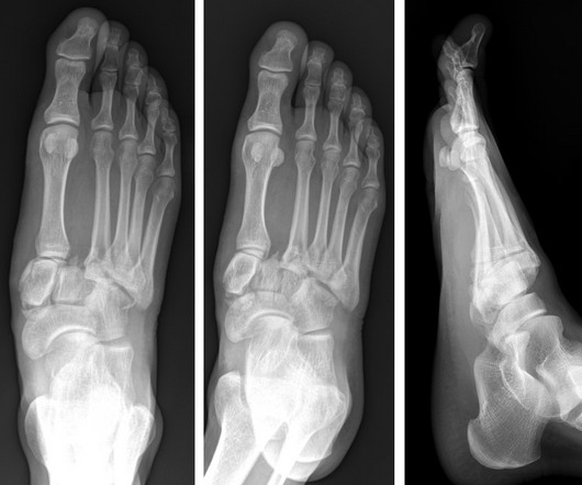
A) AP radiograph of Lisfranc Fracture Dislocation demonstrates the circled “fleck sign” or Lisfranc ligament avulsion fracture fragment. (B) C) The lateral radiograph notes with a circle, the dorsal sub dislocation of the metatarsal base. Radiographs should be repeated after two weeks to ensure surgery is unnecessary.

Insiders

Sign Up for our Newsletter

This site is protected by reCAPTCHA and the Google Privacy Policy and Terms of Service apply.

Trending Sources

article thumbnail

Type 3 Cuboid Fracture

Global Radiology CME

A) Dorsoplantar radiograph of the foot demonstrating an isolated fracture of the cuboid with possible extension into the tarsometatarsal joint. (B) B) Medial oblique radiograph of the foot demonstrating an isolated fracture of the cuboid. Radiographic evidence can support the diagnosis. Trauma in a 8 year old female.

article thumbnail

Generative AI, radiologist shortages feature in 2024 Minnies

AuntMinnie

His research interests span a gamut of topics, from 3D medical visualization, applications of AI in the early detection of cancer, and multidetector computed tomography/CT angiography development to oncologic imaging and Web-based education and training. I'm a radiographer,' " Stewart recalled. I thought, 'I'm not a teacher.

article thumbnail

Capsule Retention Following Capsule Endoscopy

Global Radiology CME

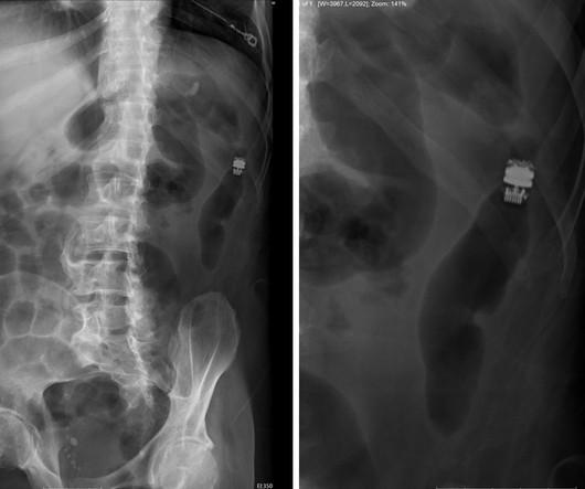
Frontal abdomen radiograph demonstrates foreign body consistent with capsule endoscopy device (pill cam) in descending colon. Computed tomography (CT) could be used to determine the capsule’s location if it is difficult to do so via x-ray. 2015.3820/2015 9. Xray of the Week Figure 1. World J Gastroenterol. 2007.06.066 8.

X-ray 52
article thumbnail

Lens Subluxation

Global Radiology CME

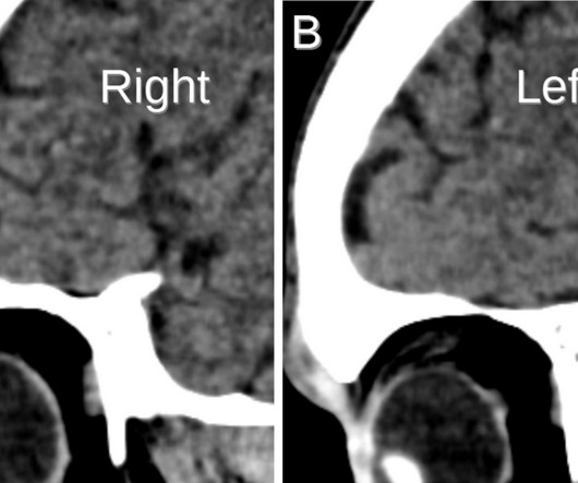
Computed tomography is also an alternative method for lens subluxation which again can show deviation of the lens (Figs. Radiographics. Anterior dislocation of the lens is serious as it may lead to angle-closure glaucoma [3,4]. Lens subluxation can be diagnosed by ultrasound which shows deviation of the lens (Fig.

article thumbnail

Ruptured Globe

Global Radiology CME

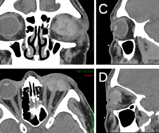
1 Imaging: Computed tomography (CT) is the recommended imaging modality for evaluating orbital trauma. Radiographics. Accuracy of Computed Tomography Imaging Criteria in the Diagnosis of Adult Open Globe Injuries by Neuroradiology and Ophthalmology. 1-2) or “flat tire” shape. Imaging of orbital trauma.