Remove 2015 Remove Diagnostic Remove Hospital Remove Radiographer
article thumbnail

Informing patients about diagnostic imaging: can we do better?

Radiology Cafe

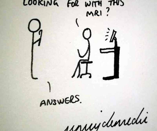
” The question was initially puzzling, as it seemed to have an obvious answer… but do we always inform patients appropriately of decisions around diagnostic imaging? Radiologists and radiographers undertaking the imaging have most likely never seen the patient before. RadioGraphics. Carlsson, S. 22(21-22):3225-3234.

article thumbnail

Lisfranc Fracture Dislocation

Global Radiology CME

A) AP radiograph of Lisfranc Fracture Dislocation demonstrates the circled “fleck sign” or Lisfranc ligament avulsion fracture fragment. (B) C) The lateral radiograph notes with a circle, the dorsal sub dislocation of the metatarsal base. Radiographs should be repeated after two weeks to ensure surgery is unnecessary.

Insiders

Sign Up for our Newsletter

This site is protected by reCAPTCHA and the Google Privacy Policy and Terms of Service apply.

Trending Sources

article thumbnail

Plastic Bowing Fracture Ulna

Global Radiology CME

Figure 2 A: AP view radiograph of right forearm. B: Lateral radiograph view of right forearm. An angulated fracture of the distal midshaft radius is also visualized, but there is also bowing of the ulna that is more appreciated on the lateral radiograph view. 8 Year Old Male With Trauma Due To A Fall. Xray of the Week Figure 1.

article thumbnail

Capsule Retention Following Capsule Endoscopy

Global Radiology CME

Frontal abdomen radiograph demonstrates foreign body consistent with capsule endoscopy device (pill cam) in descending colon. 1 ] This diagnostic procedure involves swallowing a pill-sized camera that records thousands of images of the alimentary canal including the small intestine, an area difficult to examine via traditional endoscopy.

X-ray 52
article thumbnail

Lens Dislocation

Global Radiology CME

CT is the most commonly used diagnostic modality in lens dislocation and can clearly show lens displacement [4]. Radiographics. A study of 166 hospitalized cases. Published 2015 May 27. He plans on pursuing a residency in Diagnostic Radiology. The axial CT scan above demonstrates a right lens dislocation (Fig.

article thumbnail

4 Estrategias para Reducir Errores en la Elaboración de Informes en Radiología

Everything Rad

Adrian Brady, presidente de la Sociedad Europea de Radiología y radiólogo consultor del Hospital de la Universidad de Mercy en Cork, Irlanda. Es director médico del Hereditary Haemorrhagic Telangiectasia (HHT) (con sede en el Hospital de la Universidad de Mercy). Radiographics (2015);35:1668-1676.

article thumbnail

Quatro Estratégias para Reduzir os Erros Cometidos na Geração de Relatórios na Radiologia

Everything Rad

Por: Professor Adrian Brady, presidente da European Society of Radiology e consultor em radiologia do Mercy University Hospital em Cork, na Irlanda. Ele é chefe da equipe médica do Centro Nacional Irlandês de Telangiectasia Hemorrágica Hereditária (Hereditary Haemorrhagic Telangietasia, HHT), com sede no Mercy University Hospital.