Remove 2015 Remove Imaging Modalities Remove Radiographer
article thumbnail

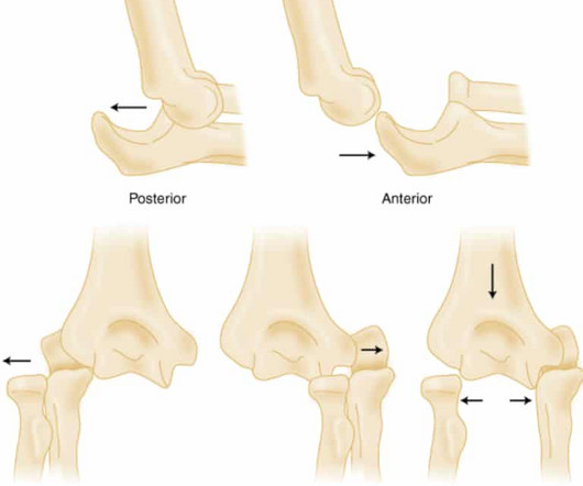
Elbow Dislocations

REBEL EM

2015 Jan;38(1):42-4. Anterior elbow dislocation with recurrent instability. Acta Orthop Belg. 2003 Apr;69(2):197-200. PMID: 12769023 Skelley NW, Chamberlain A. A novel reduction technique for elbow dislocations. Orthopedics. doi: 10.3928/01477447-20150105-05. PMID: 25611409 Stoneback JW, Owens BD, Sykes J, Athwal GS, Pointer L, Wolf JM.

article thumbnail

WBCT and its Evolving Role in OA Research and Clinical Practice

CurveBeam AI

Key Points: Weight bearing CT (WBCT) can detect signs of osteoarthritis (OA), such as osteophytes, subchondral cysts, and joint space narrowing better than radiographs. 43 (3) (2015) 213-220 ) JSW mapping has historically been performed manually, and can suffer from operator error. Segal et al.

Clinic 52
Insiders

Sign Up for our Newsletter

This site is protected by reCAPTCHA and the Google Privacy Policy and Terms of Service apply.

Trending Sources

article thumbnail

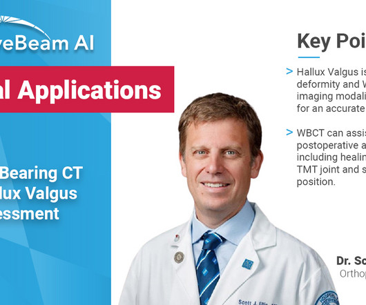
WBCT Indication Series: Hallux Valgus

CurveBeam AI

Diagnosis While radiographs are typically sufficient to make the diagnosis, WBCT scans may be useful to plan surgical treatment. Accurately assess sesamoid position as plain radiographs cannot determine whether the sesamoids have been reduced within their grooves 5. Accurately assess healing in the 1st TMT joint 4. Epub 2020 Nov 21.

article thumbnail

Ruptured Globe

Global Radiology CME

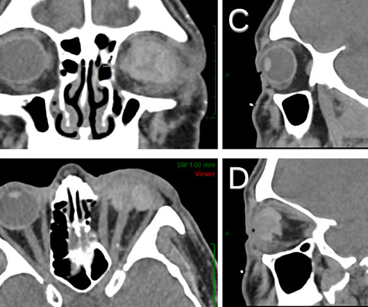
1 Imaging: Computed tomography (CT) is the recommended imaging modality for evaluating orbital trauma. Imaging of orbital trauma. Radiographics. High intraocular pressure leads to ruptures at weak areas in the eye wall, including rectus muscle insertion sites, the limbus, and the optic nerve. Clin Ophthalmol.

article thumbnail

A brief history of radiology

Radiology Cafe

after seeing the image. (2) Photoprint from radiograph by W.K. 3) In the early twentieth century, it was a common goal for investigators to try to find a way to separate the superimposed shadows that were recorded when a complex structure was shown on a radiograph. (3) 15) Radiologists needed a common means for sharing images.

article thumbnail

Emergency Guide to Stroke Neuroimaging

Northwestern EM

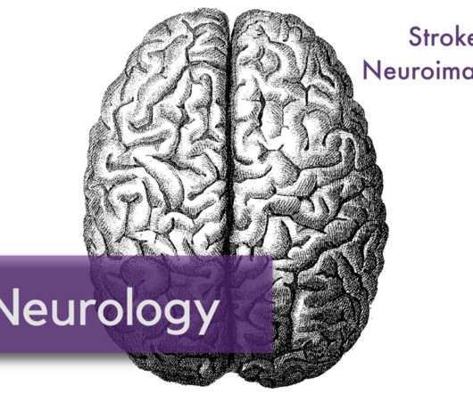
3] To identify a causative vascular lesion, which may or may not be amenable or contraindicatory to thrombolysis Non-Contrast Head CT NCCT is usually the first imaging modality obtained in the acute evaluation for stroke. Radiographics. 2015 Jul;138(Pt 7):1919-31. 2015 Mar 12;372(11):1009-18. N Engl J Med.