This site uses cookies to improve your experience. To help us insure we adhere to various privacy regulations, please select your country/region of residence. If you do not select a country, we will assume you are from the United States. Select your Cookie Settings or view our Privacy Policy and Terms of Use.
Cookie Settings
Cookies and similar technologies are used on this website for proper function of the website, for tracking performance analytics and for marketing purposes. We and some of our third-party providers may use cookie data for various purposes. Please review the cookie settings below and choose your preference.
Used for the proper function of the website
Used for monitoring website traffic and interactions
Cookie Settings
Cookies and similar technologies are used on this website for proper function of the website, for tracking performance analytics and for marketing purposes. We and some of our third-party providers may use cookie data for various purposes. Please review the cookie settings below and choose your preference.
Strictly Necessary: Used for the proper function of the website
Performance/Analytics: Used for monitoring website traffic and interactions
The Society publishes five established journals: Radiology, RadioGraphics, Radiology: Artificial Intelligence, Radiology: Cardiothoracic Imaging and Radiology: Imaging Cancer. RadioGraphics , RSNA’s premier educational journal, edited by Christine ‘Cooky’ Menias , M.D., Klein , M.D., RSNA Board Liaison for Publications.
Radiologists and radiographers undertaking the imaging have most likely never seen the patient before. The situation and the uncertainty about the coming results scared me but interaction with radiographers helped me through’: a qualitative study on patients’ experiences of magnetic resonance imaging examinations. RadioGraphics.
Figure 2 A: AP view radiograph of right forearm. B: Lateral radiograph view of right forearm. An angulated fracture of the distal midshaft radius is also visualized, but there is also bowing of the ulna that is more appreciated on the lateral radiograph view. 8 Year Old Male With Trauma Due To A Fall. Xray of the Week Figure 1.
Sites: Investigators recruited patients at 31 French emergency departments at university and nonuniversity hospitals Duration : June 1, 2009 to March 31, 2015. Practice Updates: While published in 2023, the data from this trial were gathered between 2009 and 2015. Population: Inclusion: Patients between 18–50 years of age.
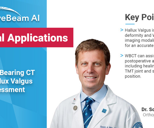
Key Points: Weight bearing CT (WBCT) can detect signs of osteoarthritis (OA), such as osteophytes, subchondral cysts, and joint space narrowing better than radiographs. 43 (3) (2015) 213-220 ) JSW mapping has historically been performed manually, and can suffer from operator error. Segal et al.
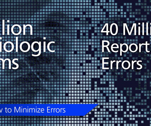
Radiographics (2015);35:1668-1676. Radiographics (2015); 35:1668-1676. American Journal of Roentgenology AJR (2007);188:1173-1178 2 Bruno MA, Walker EA, Abujudeh HH. Understanding and confronting our mistakes: the epidemiology of error in radiology and strategies for error reduction.
A) Dorsoplantar radiograph of the foot demonstrating an isolated fracture of the cuboid with possible extension into the tarsometatarsal joint. (B) B) Medial oblique radiograph of the foot demonstrating an isolated fracture of the cuboid. Radiographic evidence can support the diagnosis. Trauma in a 8 year old female.
Diagnosis While radiographs are typically sufficient to make the diagnosis, WBCT scans may be useful to plan surgical treatment. Accurately assess sesamoid position as plain radiographs cannot determine whether the sesamoids have been reduced within their grooves 5. . • Assess congruency and degenerative changes at the 1st MTP joint.
A) AP radiograph of Lisfranc Fracture Dislocation demonstrates the circled “fleck sign” or Lisfranc ligament avulsion fracture fragment. (B) C) The lateral radiograph notes with a circle, the dorsal sub dislocation of the metatarsal base. Radiographs should be repeated after two weeks to ensure surgery is unnecessary.
Frontal abdomen radiograph demonstrates foreign body consistent with capsule endoscopy device (pill cam) in descending colon. A prospective study of the utility of abdominal radiographs after capsule endoscopy for the diagnosis of capsule retention. 2015.3820/2015 9. Xray of the Week Figure 1. World J Gastroenterol.
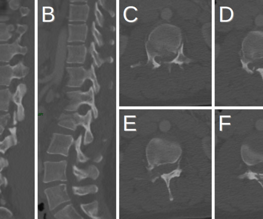
Plain radiograph shows empty vertebral body sign, which results from displacement of the spinous processes [3]. Radiographics. In 2015, Dr. Rice and Natalie Rice founded ,Global Radiology CME, to provide innovative radiology education at exciting international destinations, with the world's foremost authorities in their field.
Radiographics. Published 2015 May 27. In 2015, Dr. Rice and Natalie Rice founded Global Radiology CME to provide innovative radiology education at exciting international destinations, with the world's foremost authorities in their field. Afr J Emerg Med. 2019;9(2):106-107. doi: 10.1016/j.afjem.2019.01.001 1985;62(5):352-356.
To address this problem, Chhabra's group designed the MSKI-RADS system and assessed its efficacy using data from 208 adult patients with suspected extremity infections who underwent both x-ray and MRI exams between June 2015 and May 2019. and the team found no correlation between reader experience and overall accuracy (p = 0.94).
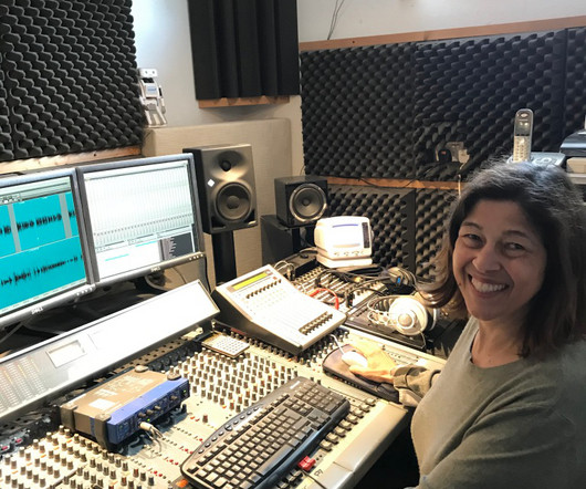
I'm a radiographer,' " Stewart recalled. My aha moment started in the fall of 2015 when I realized that informatics played such a huge role in medical imaging," Stewart said. "I I thought, 'I'm not a teacher. But Hennessy encouraged her.
JACCC Cardiovasc Interv 2015. in the paper but 2.7% to ≈0.99 (p<0.001) Mean MPI/Tei Index≈ 0.47 A prospective, Single-Arm Multicenter Trial of Ultrasound-Facilitated, Catheter-Directed, Low-Dose Fibrinolysis for Acute Massive and Submassive Pulmonary Embolism: The SEATTLE II Study. PMID: 26315743 Tapson VF et al.
Radiographics. In 2015, Dr. Rice and Natalie Rice founded ,Global Radiology CME, to provide innovative radiology education at exciting international destinations, with the world's foremost authorities in their field. 2006;10(4):345-350. doi: 10.1016/j.jaapos.2006.01.218 2006.01.218 Kubal WS. Imaging of orbital trauma. doi: 10.1148/rg.286085523
KUB indicates kidneys, ureter, and bladder (plain abdominal radiograph); CT, computed tomography; and PCD, percutaneous catheter drainage. RadioGraphics. Asterisk indicates the presence of 2 or more of the following risk factors: thrombocytopenia, acute renal failure, disturbance of consciousness, and shock. 2000;160(6):797-805.
He graduated from UMBC in 2015 with a major in Biochemistry and Molecular Biology. Seeing the radiographic images made medical education come to life for him. Jay Vora is a medical student at Edward Via College of Osteopathic Medicine (VCOM) – Virginia and plans to pursue a residency in diagnostic radiology.
Radiographics (2015);35:1668-1676. Radiographics (2015); 35:1668-1676. American Journal of Roentgenology AJR (2007);188:1173-1178 2 Bruno MA, Walker EA, Abujudeh HH. Understanding and confronting our mistakes: the epidemiology of error in radiology and strategies for error reduction.
Radiographics. In 2015, Dr. Rice and Natalie Rice founded Global Radiology CME to provide innovative radiology education at exciting international destinations, with the world's foremost authorities in their field. Clinical features of single and repeated globe rupture after penetrating keratoplasty. Clin Ophthalmol. 2013;7:461-465.
Photoprint from radiograph by W.K. 3) In the early twentieth century, it was a common goal for investigators to try to find a way to separate the superimposed shadows that were recorded when a complex structure was shown on a radiograph. (3) This is now known as ‘Hand mit Ringen’. (1) after seeing the image. (2) Röntgen, 1895.
I went through all this in 2015 when I discovered I had a Grade 1 benign posterior fossa meningioma. and to many people from the team at Addenbrooke’s, including a medical physicist and a research radiographer. A brain tumour diagnosis, like all major events, can set in place a chain of emotions, among them anger, fear and denial.
Dr Strickland (the current head of the Royal College of Radiologists) speaking in 2015 said, “Radiologists need to behave as doctors, not just imagers who issue a report and then go away without any interaction…….radiologists 2011, Radiographics, pp. Hough, Andrew. 2011, The Telegraph. Monsky, Derek S. Vien, Daniel P.
Most common organism is Staph Aureus (36-67% of cases) ( Boody 2015 ). Executive Summary: 2015 Infectious Disease Society of America (IDSA) Clinical Practice Guidelines for the Diagnosis and Treatment of Native Vertebral Osteomyelitis in Adults. Clin Infect Dis 2015 Sept 15;61(6):859-63. Other pathogens include: E.
Radiographics (2015);35:1668-1676. Radiographics (2015); 35:1668-1676. American Journal of Roentgenology AJR (2007);188:1173-1178 2 Bruno MA, Walker EA, Abujudeh HH. Understanding and confronting our mistakes: the epidemiology of error in radiology and strategies for error reduction.
Radiographics (2015); 35:1668-1676. Radiographics (2015); 35:1668-1676. American Journal of Roentgenology AJR (2007); 188:1173-1178 2 Bruno MA, Walker EA, Abujudeh HH. Understanding and confronting our mistakes: the epidemiology of error in radiology and strategies for error reduction.
Radiographics (2015); 35:1668-1676. Radiographics (2015); 35:1668-1676. American Journal of Roentgenology AJR (2007); 188:1173-1178 2 Bruno MA, Walker EA, Abujudeh HH. Understanding and confronting our mistakes: the epidemiology of error in radiology and strategies for error reduction.
It has well-defined radiographic features and various clinical presentations. They described six radiographic phenotypes of CSVD: (1) recent small subcortical infarct, (2) white matter hyperintensity, (3) lacune of presumed vascular origin, (4) widened perivascular spaces, (5) cerebral microbleed, and (6) brain atrophy.
It is important to remember that as of 2015, both IV tPA and endovascular thrombectomy are considered standard-of-care, and any patient presenting with acute ischemic stroke must undergo full workup and consideration of both treatments based upon national society / consensus guidelines. Radiographics. 2015 Jul;138(Pt 7):1919-31.
We organize all of the trending information in your field so you don't have to. Join 5,000 users and stay up to date on the latest articles your peers are reading.
You know about us, now we want to get to know you!
Let's personalize your content
Let's get even more personalized
We recognize your account from another site in our network, please click 'Send Email' below to continue with verifying your account and setting a password.


Let's personalize your content