Remove 2016 Remove Clinic Remove Diagnose Remove Global Radiology
article thumbnail

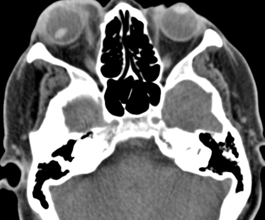
Lens Subluxation

Global Radiology CME

Lens subluxation can be diagnosed by ultrasound which shows deviation of the lens (Fig. Trauma-Induced Bilateral Ectopia Lentis Diagnosed with Point-of-Care Ultrasound. 2015.01.004 Sai Kilaru is a medical student at Central Michigan University College of Medicine and plans to pursue a residency in diagnostic radiology.

article thumbnail

Lens Dislocation

Global Radiology CME

Ultrasound has been shown to rapidly diagnose lens dislocation as it can visualize internal structures of the globe [2, 7]. He plans on pursuing a residency in Diagnostic Radiology. Austin discovered his passion for radiology during his clinical rotations and finds using imaging to diagnose patients very rewarding.

Insiders

Sign Up for our Newsletter

This site is protected by reCAPTCHA and the Google Privacy Policy and Terms of Service apply.

Trending Sources

article thumbnail

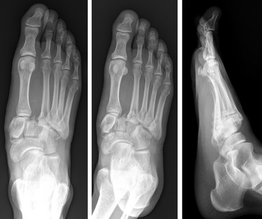
Lisfranc Fracture Dislocation

Global Radiology CME

of all diagnosed fractures. With over 20% of Lisfranc fractures missed upon presentation, it is important to diagnose these injuries promptly, as lack of diagnosis could lead to future foot deformities, midfoot arthritis, pain, chronic instability, and disability [1,5]. All posts by Rebeca Santos Kevin M.

article thumbnail

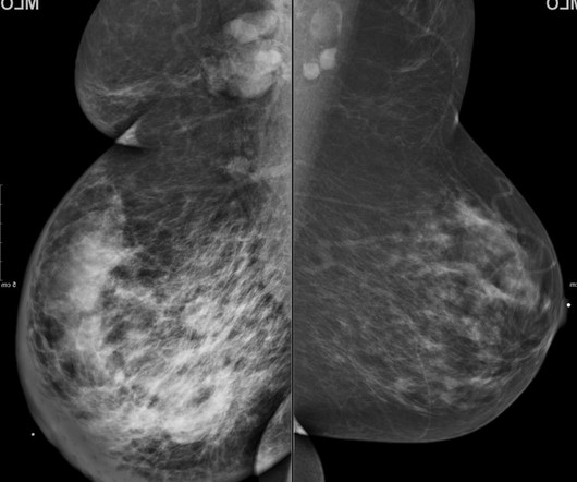
Inflammatory Breast Carcinoma

Global Radiology CME

Discussion: 63-year-old woman presented to a clinic with a one month history of progressively enlarging right breast with redness and tenderness. Both tissue diagnosis of malignancy and clinical evidence of inflammatory disease are required to confirm the diagnosis of IBC [3].

article thumbnail

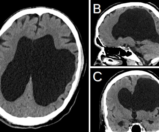
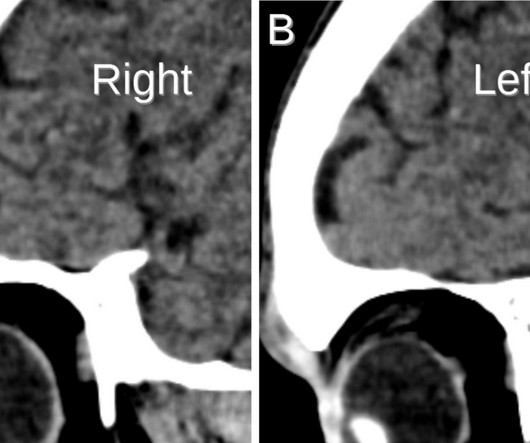
Porencephaly

Global Radiology CME

Porencephaly is readily diagnosed via imaging. The expanding phenotype of COL4A1 and COL4A2 mutations: clinical data on 13 newly identified families and a review of the literature. Diagnostic Imaging: Obstetrics, Elsevier, 2016, pp. He discovered his passion for radiology during the first radiology lecture at VCOM.

article thumbnail

Left Atrial Myxoma Presenting with Splenic Infarct

Global Radiology CME

Younger patients ( Clinical symptoms can be caused directly by the tumor from obstruction or in form of embolization. If a stalk or pedunculated mass is not visualized on the echo, MRI can be used to diagnose CM's and help differentiate from thrombus or pseudotumor [3].