Remove 2016 Remove Clinic Remove Diagnose Remove Ultrasound
article thumbnail

Surprising Improved Productivity with Ultrasound Worksheets

Imorgon Medical

” In this article, I discuss the importance of both measurement transfer and electronic ultrasound worksheets in ultrasound reporting software to achieve these efficiency gains. Ultrasound worksheet templates serve as checklists for sonographers, enhancing the quality and consistency of observations during exams.

Insiders

Sign Up for our Newsletter

This site is protected by reCAPTCHA and the Google Privacy Policy and Terms of Service apply.

Trending Sources

article thumbnail

Lens Subluxation

Global Radiology CME

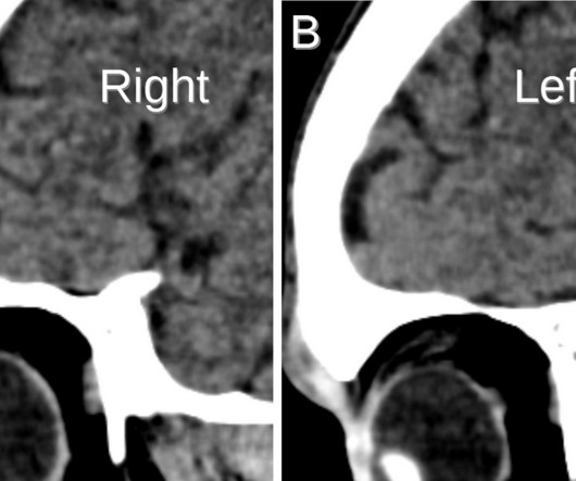
A: Ultrasound showing deviation of the right lens margin posteriorly into the anechoic vitreous humor (red arrow). B: Ultrasound demonstrates the normal location of the left lens in the iris (green arrow). Lens subluxation can be diagnosed by ultrasound which shows deviation of the lens (Fig. Radiographics. J Emerg Med.

article thumbnail

Lens Dislocation

Global Radiology CME

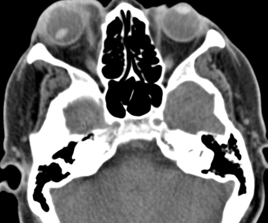
Ultrasound has been shown to rapidly diagnose lens dislocation as it can visualize internal structures of the globe [2, 7]. Austin discovered his passion for radiology during his clinical rotations and finds using imaging to diagnose patients very rewarding. He plans on pursuing a residency in Diagnostic Radiology.

article thumbnail

Inflammatory Breast Carcinoma

Global Radiology CME

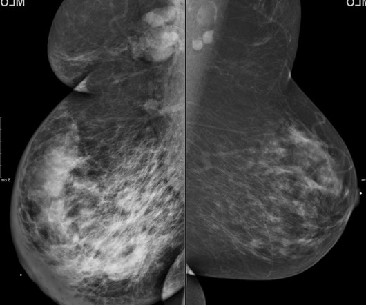
Discussion: 63-year-old woman presented to a clinic with a one month history of progressively enlarging right breast with redness and tenderness. Ultrasound guided biopsy yielded invasive ductal carcinoma with lymphatic invasion. However, if the lesion is not visualized on imaging, this does not preclude the diagnosis of IBC [1].

article thumbnail

Porencephaly

Global Radiology CME

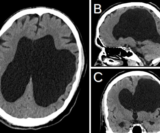
Porencephaly is readily diagnosed via imaging. Antenatal ultrasound may show one or more intracranial cysts that communicate with the ventricular system and/or subarachnoid space. The expanding phenotype of COL4A1 and COL4A2 mutations: clinical data on 13 newly identified families and a review of the literature. 56 (2): 287–290.

article thumbnail

Why understanding breast density matters

The British Institute of Radiology

1) and may go undetected until they are larger and more likely to present with clinical symptoms [4]. In these women, screening tests, such as ultrasound or MRI, when added to mammography, substantially increase the detection of early-stage breast cancer. Mammograms will miss about 40% of cancers in women with extremely dense breasts.