Remove 2016 Remove Diagnose Remove Mammogram Remove Ultrasound
article thumbnail

Abbreviated MRI suitable in subsequent screening of dense breasts

AuntMinnie

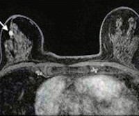
(B) Axial subtracted contrast-enhanced fat-suppressed T1-weighted image from a subsequent-round abbreviated MRI examination performed two years later shows a new 5-mm enhancing mass in the upper outer right breast (arrow), which was not seen on a mammogram performed five months prior. The exam was assessed as BI-RADS category 5. PPV2 21.3%

MRI 111
article thumbnail

USPSTF finalizes breast cancer screening recommendations

AuntMinnie

What’s more, the USPSTF concluded that there was insufficient evidence to recommend supplemental screening with MRI or ultrasound in women, regardless of breast density. Furthermore, these women should also begin annual mammography at ages 25 to 40.

Insurance 102
Insiders

Sign Up for our Newsletter

This site is protected by reCAPTCHA and the Google Privacy Policy and Terms of Service apply.

Trending Sources

article thumbnail

Why understanding breast density matters

The British Institute of Radiology

Women with dense breasts are BOTH more likely to develop breast cancer and more likely to have that cancer missed on a mammogram [5] Fig. 1 – Cancer on a mammogram of a fatty vs a dense breast What is Dense Breast Tissue? Breast density is determined through a mammogram and described as one of four categories (Fig.

article thumbnail

Inflammatory Breast Carcinoma

Global Radiology CME

A) Mammogram MLO view. Mammogram CC view. A mammogram demonstrated focal asymmetries involving most of the anterior and mid right breast with diffuse skin thickening, trabecular coarsening, increased overall density, and enlarged right axillary lymph nodes. What is the diagnosis? Xray of the Week Figure 1.