article thumbnail

Chalk Stick Fracture in Ankylosing Spondylitis

Global Radiology CME

Ankylosing Spondylitis: Ankylosing spondylitis (AS) is a seronegative spondyloarthropathy and chronic inflammatory disease of the axial skeleton that leads to the partial or complete fusion and rigidity of the spine. Because radiography may not detect early signs of AS, non-enhanced MRI is recommended in identifying early signs of disease.

article thumbnail

Capsule Retention Following Capsule Endoscopy

Global Radiology CME

Capsule Retention and Risk Factors: Capsule endoscopy is used for evaluating small-bowel disorders, such as bleeds and Crohn disease.[ For patients with inflammatory bowel disease, the use of steroids has been shown to assist the excretion of capsules in up to 20-30% of all CR cases.[

X-ray 52
Insiders

Sign Up for our Newsletter

This site is protected by reCAPTCHA and the Google Privacy Policy and Terms of Service apply.

Trending Sources

article thumbnail

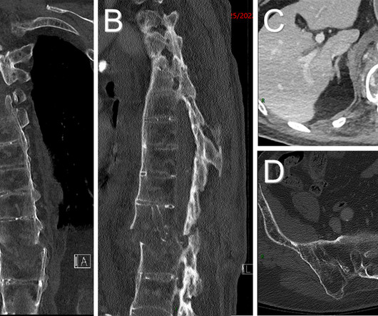
Type 3 Cuboid Fracture

Global Radiology CME

Cuboid Fracture Classification: In 2016, Fenton et al. Malunion, degenerative joint disease, persistent subluxation, and prolonged pain are complications of mismanaged cuboid fractures [2]. Rice's passion for state of the art radiology and teaching includes acting as a guest lecturer at UCLA.

article thumbnail

Tricuspid Valve Repair with MitraClip

Global Radiology CME

Rarer causes include rheumatic disease, congenital, myxomatous degeneration, endocarditis, or pulmonary hypertension [3]. Diagnosis and treatment of tricuspid valve disease: current and future perspectives. Rice's passion for state of the art radiology and teaching includes acting as a guest lecturer at UCLA. Circulation.

article thumbnail

Acute Infective Vasitis

Global Radiology CME

Discussion: Vasitis refers to a rare inflammatory disease of the vas deferens (1). Rice, MD is the president of Global Radiology CME and is a radiologist with Cape Radiology Group. Rice's passion for state of the art radiology and teaching includes acting as a guest lecturer at UCLA.

article thumbnail

Inflammatory Breast Carcinoma

Global Radiology CME

Both tissue diagnosis of malignancy and clinical evidence of inflammatory disease are required to confirm the diagnosis of IBC [3]. Around 3-6 months and most commonly within 3 months into the disease progression, changes of the skin and development of an underlying mass will be appreciated and are used to validate the diagnosis.

article thumbnail

Porencephaly

Global Radiology CME

Avery’s Diseases of the Newborn, Elsevier, 2018, pp. Diagnostic Imaging: Obstetrics, Elsevier, 2016, pp. Jay Vora is a medical student at Edward Via College of Osteopathic Medicine (VCOM) – Virginia and plans to pursue a residency in diagnostic radiology. Am J Dis Child. 1991;145(6):688-695. doi: 10.1001/archpedi.1991.02160060106030