Remove 2016 Remove Disease Remove Radiographer
article thumbnail

WBCT Indication Series: Charcot Arthropathy

CurveBeam AI

Additionally, in the early stages of Charcot (Eichenholtz stages 0 and 1), a WBCT scan at regular intervals may be used to track progression and instability of the disease process.​ Non-WB radiographs could not determine instability or severity of midfoot collapse; surgical intervention postponed. 2016, October). Diabet Med.

article thumbnail

Meet the Minnies 2024 finalists

AuntMinnie

His research interests include using structural and functional MRI -- particularly ultrahigh-field, 7-tesla MRI -- to map brain microstructure and develop neurosurgical treatment of brain tumors, epilepsy, and neurodegenerative and movement disorders such as Parkinson's disease, essential tremor, and dystonia. Perry Pickhardt, MD.

Insiders

Sign Up for our Newsletter

This site is protected by reCAPTCHA and the Google Privacy Policy and Terms of Service apply.

Trending Sources

article thumbnail

REBEL Cast Ep123: Reduced-Dose Systemic Peripheral Alteplase in Massive PE?

REBEL EM

in the paper but 2.7% to ≈0.99 (p<0.001) Mean MPI/Tei Index≈ 0.47 selection bias) Unclear therapies prior to lytics (i.e. Thrombolysis Compared with Heparin for the Initial Treatment of Pulmonary Embolism: A Meta-Analysis of the Randomized Controlled Trials. PMID: 15262836 Sharifi M et al. PMID: 27422214 Wang C et al.

Hospital 136
article thumbnail

Type 3 Cuboid Fracture

Global Radiology CME

A) Dorsoplantar radiograph of the foot demonstrating an isolated fracture of the cuboid with possible extension into the tarsometatarsal joint. (B) B) Medial oblique radiograph of the foot demonstrating an isolated fracture of the cuboid. Radiographic evidence can support the diagnosis. Trauma in a 8 year old female.

article thumbnail

Minnies winners for 2023 highlight AI, innovation in radiology

AuntMinnie

He received his first Most Influential Radiology Researcher Minnie in 2016, largely due to his previous work in CT colonography. We should be leveraging the ‘free’ body composition data that is available on all abdominal CTs.”— Pickhardt graduated from UW in 1991 with a Bachelor of Science in physics.

Radiology 132
article thumbnail

The radiologist in 22 years time

Radiology Cafe

It is the merging of imaging physics and disease biology that will allow radiologists to interpret images in a clinical context and so help guide clinicians, with correspondingly less involvement in only a single phase of the process. 2016, International Journal of scientific research, pp. 2016, Nature. 2016, The Economist.

article thumbnail

Capsule Retention Following Capsule Endoscopy

Global Radiology CME

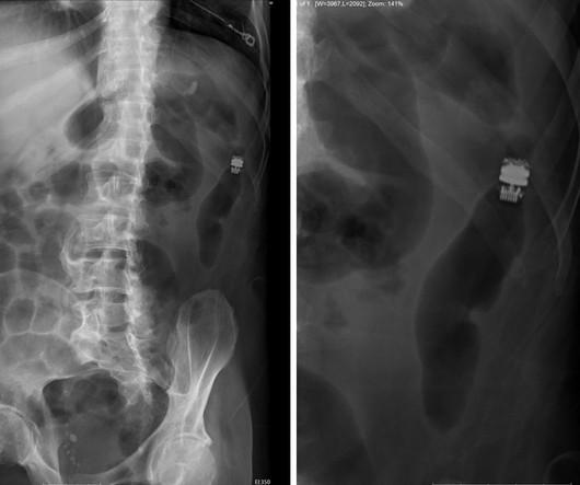
Frontal abdomen radiograph demonstrates foreign body consistent with capsule endoscopy device (pill cam) in descending colon. Capsule Retention and Risk Factors: Capsule endoscopy is used for evaluating small-bowel disorders, such as bleeds and Crohn disease.[ What are the important findings in this case. World J Gastroenterol.

X-ray 52