Remove 2016 Remove Global Radiology Remove Radiologist Remove Ultrasound
article thumbnail

Acute Infective Vasitis

Global Radiology CME

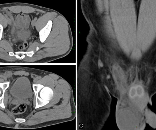
Ultrasound of acute vasitis shows a heterogenous and thickened vas deferens. Color Doppler ultrasound of the same section as Figure 4. With ultrasound, acute vasitis shows a heterogenous, hypoechoic and thickened vas deferens with hyperemia on color Doppler (3-6)(Figs. Journal of Ultrasound in Medicine. J Ultrasound Med.

article thumbnail

Lens Subluxation

Global Radiology CME

A: Ultrasound showing deviation of the right lens margin posteriorly into the anechoic vitreous humor (red arrow). B: Ultrasound demonstrates the normal location of the left lens in the iris (green arrow). Lens subluxation can be diagnosed by ultrasound which shows deviation of the lens (Fig. Radiographics. 2008;28(6):1729-1739.

Insiders

Sign Up for our Newsletter

This site is protected by reCAPTCHA and the Google Privacy Policy and Terms of Service apply.

Trending Sources

article thumbnail

Inflammatory Breast Carcinoma

Global Radiology CME

Ultrasound guided biopsy yielded invasive ductal carcinoma with lymphatic invasion. Once the clinical picture and baseline laboratory tests for tumor markers like cancer antigen 15-3 and CEA confirm the diagnosis, local imaging with mammogram and ultrasound will help guide management as standards of care [1]. Breast Cancer.

article thumbnail

Porencephaly

Global Radiology CME

Antenatal ultrasound may show one or more intracranial cysts that communicate with the ventricular system and/or subarachnoid space. Ultrasound and Magnetic Resonance in Prenatal Diagnosis of Congenital Anomalies.” Diagnostic Imaging: Obstetrics, Elsevier, 2016, pp. Porencephaly is readily diagnosed via imaging.

article thumbnail

Lens Dislocation

Global Radiology CME

Ultrasound has been shown to rapidly diagnose lens dislocation as it can visualize internal structures of the globe [2, 7]. Rice's passion for state of the art radiology and teaching includes acting as a guest lecturer at UCLA. The axial CT scan above demonstrates a right lens dislocation (Fig.