article thumbnail

Lisfranc Fracture Dislocation

Global Radiology CME

A) AP radiograph of Lisfranc Fracture Dislocation demonstrates the circled “fleck sign” or Lisfranc ligament avulsion fracture fragment. (B) C) The lateral radiograph notes with a circle, the dorsal sub dislocation of the metatarsal base. Radiographs should be repeated after two weeks to ensure surgery is unnecessary.

article thumbnail

Type 3 Cuboid Fracture

Global Radiology CME

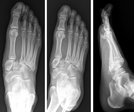
A) Dorsoplantar radiograph of the foot demonstrating an isolated fracture of the cuboid with possible extension into the tarsometatarsal joint. (B) B) Medial oblique radiograph of the foot demonstrating an isolated fracture of the cuboid. Radiographic evidence can support the diagnosis. Trauma in a 8 year old female.

Insiders

Sign Up for our Newsletter

This site is protected by reCAPTCHA and the Google Privacy Policy and Terms of Service apply.

Trending Sources

article thumbnail

The radiologist in 22 years time

Radiology Cafe

If this is where we are at already, it is not unreasonable to imagine that in 23 years AI will be capable of reporting imaging studies (CT or magnetic resonance imaging (MRI)) with a higher accuracy than humans and that these reports will no longer be routinely reported by human radiologists out of hours. 2016, Nature.

article thumbnail

Emergency Guide to Stroke Neuroimaging

Northwestern EM

Magnetic resonance imaging and computed tomography in emergency assessment of patients with suspected acute stroke: a prospective comparison. Radiographics. Approach to reperfusion therapy for acute ischemic stroke. Last Update: September 14, 2018. Accessed from [link] Chaela JA, Kidwell CS, Nentwich LM, et al.