Remove 2016 Remove Magnetic Resonance Imaging Remove Radiographer Remove Radiologist
article thumbnail

Type 3 Cuboid Fracture

Global Radiology CME

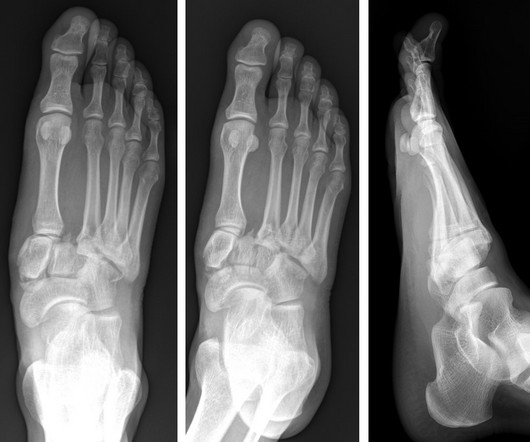
A) Dorsoplantar radiograph of the foot demonstrating an isolated fracture of the cuboid with possible extension into the tarsometatarsal joint. (B) B) Medial oblique radiograph of the foot demonstrating an isolated fracture of the cuboid. Radiographic evidence can support the diagnosis. Trauma in a 8 year old female.

article thumbnail

Lisfranc Fracture Dislocation

Global Radiology CME

A) AP radiograph of Lisfranc Fracture Dislocation demonstrates the circled “fleck sign” or Lisfranc ligament avulsion fracture fragment. (B) C) The lateral radiograph notes with a circle, the dorsal sub dislocation of the metatarsal base. Radiographs should be repeated after two weeks to ensure surgery is unnecessary.

article thumbnail

The radiologist in 22 years time

Radiology Cafe

I believe that the first main difference seen in the radiologists role in 2040 will be the change that artificial intelligence (AI) will have made on the diagnostic role that radiologists currently perform. There are several protective factors, the first being that radiologists do much more than just report scans.