This site uses cookies to improve your experience. To help us insure we adhere to various privacy regulations, please select your country/region of residence. If you do not select a country, we will assume you are from the United States. Select your Cookie Settings or view our Privacy Policy and Terms of Use.
Cookie Settings
Cookies and similar technologies are used on this website for proper function of the website, for tracking performance analytics and for marketing purposes. We and some of our third-party providers may use cookie data for various purposes. Please review the cookie settings below and choose your preference.
Used for the proper function of the website
Used for monitoring website traffic and interactions
Cookie Settings
Cookies and similar technologies are used on this website for proper function of the website, for tracking performance analytics and for marketing purposes. We and some of our third-party providers may use cookie data for various purposes. Please review the cookie settings below and choose your preference.
Strictly Necessary: Used for the proper function of the website
Performance/Analytics: Used for monitoring website traffic and interactions
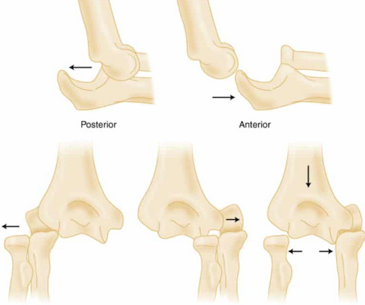
Elbow Dislocation Definition: Disarticulation of the proximal radius & ulna bones from the humerus Epidemiology: Incidence Second most common joint dislocation (after shoulder) in adults Most commonly dislocated joint in children Accounts for 10-25% of all injuries to the elbow ( Cohen 1998 ) Posterolateral is the most common type of dislocation (..)
Perry Pickhardt, MD, University of Wisconsin -- Madison Turning up in the category of Most Influential Radiology Researcher isn't new for Perry Pickhardt, MD -- he won the category last year and in 2016. Perry Pickhardt, MD. Elliot Fishman, MD. Wagner Graduate School of Public Service in New York, NY.
He received his first Most Influential Radiology Researcher Minnie in 2016, largely due to his previous work in CT colonography. “We plan to collaborate with our clinicians in areas of preventive cardiology, endocrinology, and hepatology, for example,” Pickhardt said. Pickhardt graduated from UW in 1991 with a Bachelor of Science in physics.
Non-WB radiographs could not determine instability or severity of midfoot collapse; surgical intervention postponed. One year later, deformity had significantly progressed, but the remaining bone stock was unclear from plain radiographs. WB radiographs of the left foot demonstrated Charcot arthropathy. 2016, October).
She was the NCI and ACRIN representative on the Gynecologic Cancer International Trials Group from 2009 to 2016. She has served on the RSNA Genitourinary Scientific Program Committee and RadioGraphics Genitourinary Review Panel. She also served as chair of radiomics trials for ECOG-ACRIN.
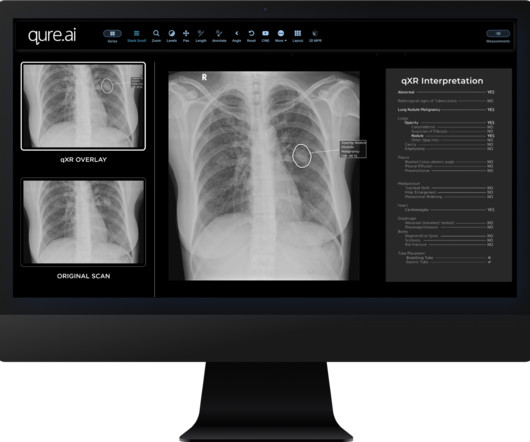
As a Chief Medical Officer, rapid pneumothorax identification adds a layer of safety to the hospital by providing a check to identify one of the most time-sensitive radiographic findings quickly.” More information: www.qure.ai Reference: [link] Related content: AI Essentials in Radiology: Experts Weigh In Qure.ai
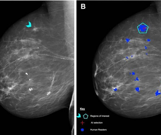
The researchers compared the AI test scores with the scores of the 552 human readers, including 315 (57%) board-certified radiologists and 237 non-radiologist readers consisting of 206 radiographers and 31 breast clinicians.
Enhancing radiographers' performance Featured in European Radiology , a second peer-reviewed study investigates whether active use of Volpara Analytics software could help breast radiographers to improve their screening image quality.
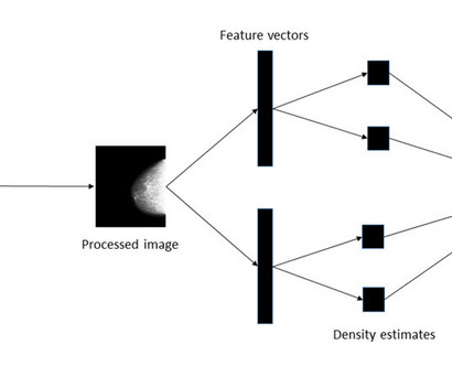
Using nearly 160,000 full-field digital mammogram images that were assigned density values on a visual analogue scale by experts (radiologists, advanced practitioner radiographers, and breast physicians) from 39,357 women, the researchers developed a procedure for estimating the density score for each mammogram image.
Figure 2 A: AP view radiograph of right forearm. B: Lateral radiograph view of right forearm. An angulated fracture of the distal midshaft radius is also visualized, but there is also bowing of the ulna that is more appreciated on the lateral radiograph view. 8 Year Old Male With Trauma Due To A Fall. Xray of the Week Figure 1.
A) Dorsoplantar radiograph of the foot demonstrating an isolated fracture of the cuboid with possible extension into the tarsometatarsal joint. (B) B) Medial oblique radiograph of the foot demonstrating an isolated fracture of the cuboid. Radiographic evidence can support the diagnosis. Trauma in a 8 year old female.
through a Govt tender (State Health and family welfare society, Govt of Tripura), which went live on September 15th, 2016. To date, 19 hospitals of Tripura have benefitted from Teleradiology services resulting in a total read-out of 61,660 radiographs by radiologists situated in Bangalore, Hyderabad, New Delhi, and other cities.
A) AP radiograph of Lisfranc Fracture Dislocation demonstrates the circled “fleck sign” or Lisfranc ligament avulsion fracture fragment. (B) C) The lateral radiograph notes with a circle, the dorsal sub dislocation of the metatarsal base. Radiographs should be repeated after two weeks to ensure surgery is unnecessary.
Plain radiograph shows empty vertebral body sign, which results from displacement of the spinous processes [3]. Radiographics. In 2016, Dr. Rice was nominated and became a semifinalist for a ,"Minnie" Award for the Most Effective Radiology Educator. Thoracolumbar Spine Injury at CT: Trauma/Emergency Radiology. doi:10.1148/rg.2016160058
Radiographics. In 2016, Dr. Rice was nominated and became a semifinalist for a ,"Minnie" Award for the Most Effective Radiology Educator. Anterior axial lens subluxation, progressive myopia, and angle closure glaucoma: recognition and treatment of atypical presentation of ectopia lentis. 2006;10(4):345-350. doi: 10.1016/j.jaapos.2006.01.218
Frontal abdomen radiograph demonstrates foreign body consistent with capsule endoscopy device (pill cam) in descending colon. A prospective study of the utility of abdominal radiographs after capsule endoscopy for the diagnosis of capsule retention. 61-year-old male with abdominal pain 15 days after capsule endoscopy. doi: 10.3748/wjg.15.2401
Radiographics. In 2016, Dr. Rice was nominated and became a semifinalist for a ,"Minnie" Award for the Most Effective Radiology Educator. Afr J Emerg Med. 2019;9(2):106-107. doi: 10.1016/j.afjem.2019.01.001 2019.01.001 Bass LJ, Potter JW. A case of spontaneous dislocated lenses. Am J Optom Physiol Opt. 1985;62(5):352-356.
A growing number of medical organisations link to the DenseBreast-info.org website, including the EFRS (European Federation of Radiographer Societies) and the Society of Radiographers. Figure 4 (b) The website includes breast screening guidelines in Europe. A comparative analysis table summarises the guidelines in each country.
Radiographics. In 2016, Dr. Rice was nominated and became a semifinalist for a "Minnie" Award for the Most Effective Radiology Educator. Clinical features of single and repeated globe rupture after penetrating keratoplasty. Clin Ophthalmol. 2013;7:461-465. doi:10.2147/OPTH.S42117 Kubal WS. Imaging of orbital trauma. doi:10.1148/rg.286085523
This study builds on data from previous studies in determining if a therapeutic approach is more broadly applicable A well-defined, and standardized radiographic scoring system was used to identify appropriate patients The study’s inclusion criteria limited the enrollment of patients who were at higher risk for complications from diverticulitis.
Geoffrey Hinton, a key pioneer in deep learning, famously remarked in 2016 that we should stop training radiologists because machine learning might soon outperform them at image interpretation, with a time frame of five to 10 years. RadioGraphics. American College of Radiology. ACR Commission on Human Resources Workforce Survey.
RadioGraphics. American College of Radiology. ACR Commission on Human Resources Workforce Survey. Harvey HB, et al. The Future of Radiology: Artificial Intelligence and Advanced Technologies. 2023;43(3):e230017. Society of Interventional Radiology. Annual Workforce Survey Report. Bureau of Labor Statistics. Radiology: Artificial Intelligence.
KUB indicates kidneys, ureter, and bladder (plain abdominal radiograph); CT, computed tomography; and PCD, percutaneous catheter drainage. RadioGraphics. In 2016, Dr. Rice was nominated and became a semifinalist for a ,"Minnie" Award for the Most Effective Radiology Educator. 2000;160(6):797-805. doi:10.1001/archinte.160.6.797
Diagnostic Imaging: Obstetrics, Elsevier, 2016, pp. Seeing the radiographic images made medical education come to life for him. In 2016, Dr. Rice was nominated and became a semifinalist for a ,"Minnie" Award for the Most Effective Radiology Educator. Textbook of Fetal Abnormalities, Elsevier, 2007, pp.
2016, International Journal of scientific research, pp. 2016, Nature. 2016, The Economist. 2016, Science Translational Medicine. 2011, Radiographics, pp. Dethe, Varsha S. Shevatkar, R.P. Discrimination of Breast Cancer with Microcalcifications on Mammography by Deep Learning. Automation and anxiety. Hough, Andrew.
in the paper but 2.7% to ≈0.99 (p<0.001) Mean MPI/Tei Index≈ 0.47 Thrombolysis Compared with Heparin for the Initial Treatment of Pulmonary Embolism: A Meta-Analysis of the Randomized Controlled Trials. PMID: 15262836 Sharifi M et al. Pulseless Electrical Activity in Pulmonary Embolism Treated with Thrombolysis (from the “PEAPETT” Study).
The synthetic control arm was obtained retrospectively from one of three acute care hospitals in the Hartford Healthcare network between 12/1/2016 and 8/30/2020. POPULATION Inclusions: Age ≥ 18 years of age Radiographically confirmed acute spontaneous or traumatic intracranial hemorrhage (i.e.
Saline load test Has mainly been supplanted by CT scan due to ease in obtaining, reported performance characteristics, consultant recommendation and difficulty in interpreting test. cefazolin or cefuroxime) If risk factors for MRSA present, use agent with activity against MRSA (i.e.
Radiographics. “Vascular Supply of the Brain and Spinal Cord” In Gray's clinical neuroanatomy: The anatomic basis for clinical neuroscience. Philadelphia, PA: Elsevier/Saunders. Mtui E, Gruener G, Dockery P. Blood Supply of the Brain.” In Fitzgerald’s Clinical Neuroanatomy and Neuroscience. Edinburgh: Elsevier Saunders. Neurosurgery.
We organize all of the trending information in your field so you don't have to. Join 5,000 users and stay up to date on the latest articles your peers are reading.
You know about us, now we want to get to know you!
Let's personalize your content
Let's get even more personalized
We recognize your account from another site in our network, please click 'Send Email' below to continue with verifying your account and setting a password.


Let's personalize your content