

Meet the Minnies 2024 finalists
AuntMinnie
OCTOBER 25, 2024
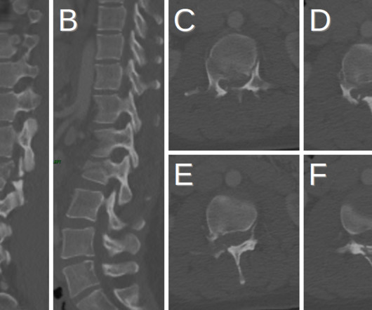
Perry Pickhardt, MD, University of Wisconsin -- Madison Turning up in the category of Most Influential Radiology Researcher isn't new for Perry Pickhardt, MD -- he won the category last year and in 2016. Perry Pickhardt, MD. Elliot Fishman, MD. Wagner Graduate School of Public Service in New York, NY.


Let's personalize your content