This site uses cookies to improve your experience. To help us insure we adhere to various privacy regulations, please select your country/region of residence. If you do not select a country, we will assume you are from the United States. Select your Cookie Settings or view our Privacy Policy and Terms of Use.
Cookie Settings
Cookies and similar technologies are used on this website for proper function of the website, for tracking performance analytics and for marketing purposes. We and some of our third-party providers may use cookie data for various purposes. Please review the cookie settings below and choose your preference.
Used for the proper function of the website
Used for monitoring website traffic and interactions
Cookie Settings
Cookies and similar technologies are used on this website for proper function of the website, for tracking performance analytics and for marketing purposes. We and some of our third-party providers may use cookie data for various purposes. Please review the cookie settings below and choose your preference.
Strictly Necessary: Used for the proper function of the website
Performance/Analytics: Used for monitoring website traffic and interactions
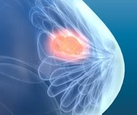
Detecting breast arterial calcifications on routine mammograms could identify women at a higher risk of future cardiovascular disease (CVD), a study published March 13 in Clinical Imaging found. Breast arterial calcifications are incidental findings on mammograms.
Patients underwent cryoablation between January 2017 and March 2023. Follow-up imaging was performed after the procedure by mammogram, ultrasound, or in some cases contrast-enhanced mammogram or MRI, based on patient eligibility and preference. cm who were poor surgical candidates or who refused surgery.
The final analysis included data collected between 2005 and 2017 from 3,529,825 screening mammograms. Women who were recalled for additional imaging, short-interval follow-up recommendations, or biopsy recommendations were less likely to return to regular screening compared with women whose mammograms were deemed to be true-negative.
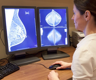
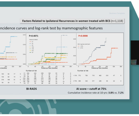
A team led by Julie Hamzah, MBBS, from Singapore General Hospital, found that symptomatic first breast cancers, dense breasts, and the presence of trabecular thickening on mammography are tied to mammogram detection failure of ipsilateral second breast cancers.
The multicenter, retrospective study included data collected between 2012 and 2017 from 1,740 women with an average of 51.5 The researchers analyzed preoperative routine mammograms via a commercially available AI algorithm (Lunit Insight MMG, Lunit ). years who were treated for DCIS.
Saige-Dx is intended to be used on women 35 years old and older, and it is not intended to replace a physician's own review of a mammogram. was created in 2017 after DeepHealth LLC was founded in 2015, and that’s when the Saige AI technology was first initiated. DeepHealth Inc.
The study included data collected between 2017 and 2019 from 140 women who underwent both modalities at 10 centers as part of a prospective case collection registry.
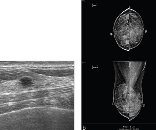
Why Breast Density Matters in Cancer Screening Dense breast tissue affects screening in two key ways: Reduced Visibility : Dense tissue appears white on mammograms, as do tumors, making it harder to detect abnormalities. Inter-radiologist Variation : Assessments can vary up to 33% 1 when different radiologists interpret the same mammograms.
This AJR accepted manuscript included 1,325 women (mean age, 53 years) with dense breasts who underwent both screening mammography and supplementary breast US within a 1-month interval from January 2017 to December 2017; prior mammogram and US were available to compare in 91.2% and 91.8%, respectively. s analysis.
From January 1, 2014 to December 31, 2016, patients temporarily discontinued all AT for 5 days before CNB; from January 1, 2017 to December 31, 2019, the cohort maintained AT during CNB.
Breast Density on a Mammogram. VIDEO “Big Concerns Remain for MRI Gadolinium Contrast Safety at RSNA 2017,” An interview with Emanuel Kanal, M.D. Ghaderi, Jordana Phillips, Hannah Perry, Parisa Lotfi, and Tejas S. Contrast-enhanced Mammography: Current Applications and Future Directions. RadioGraphics 2019 39:7, 1907-1920.
This AJR accepted manuscript included 1,325 women (mean age, 53 years) with dense breasts who underwent both screening mammography and supplementary breast US within a 1-month interval from January 2017 to December 2017; prior mammogram and US were available to compare in 91.2% and 91.8%, respectively. s analysis.
AJR:209, December 2017 ) Folds in the IMF are visualized as follows: FFDM 35% and with DBT 45% of the time. ( AJR:209, December 2017 ) What defines the visualization of the IMF?? 1993), it was shown that only 67% of mammograms met the “criteria”of a good mammogram. American Journal of Roentgenology (AJR) 2017.
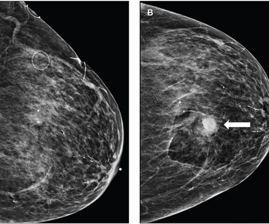
Almost 43% of women over 40 years old have dense breast tissue that can obscure lesions on traditional 2D mammograms, making cancers harder to detect and recalls more likely [6]. Women with very dense breasts have a four to five times greater risk of developing breast cancer in comparison to women with less dense breasts [7]. Baird, Martha B.
The women were diagnosed with ER+ or HER2-negative breast cancer from 2010 to 2017 and enrolled in fee-for-service Medicare Parts A and B from five years prior to diagnosis to through one year after diagnosis. The study used linked Surveillance, Epidemiology, and End Results (SEER) Medicare data of women ages 70 or older.
Cancer experts, however, have expressed concerns over the potential number of women who delayed breast cancer screenings and mammograms due to covid-related closures or backlogs. Let’s look at some of the recent numbers and revisit the importance of breast cancer screenings and mammograms. Why is Your Yearly Mammogram Important?
A deep-learning algorithm can rule out the presence of breast cancer on screening mammograms, improving specificity and yielding significant workflow and downstream savings, according to research published April 10 in Radiology. dataset 1: 143,593 mammograms interpreted by 11 breast radiologists from 2008 to 2017 U.S.
AI is currently being used for many applications in radiology; for example, in speech recognition, detecting and characterising lung nodules, characterising liver lesions in MRI and prioritizing follow up evaluations and identifying and characterizing microcalcifications in mammograms. (21) Prevedello. Radiol Bras.
We organize all of the trending information in your field so you don't have to. Join 5,000 users and stay up to date on the latest articles your peers are reading.
You know about us, now we want to get to know you!
Let's personalize your content
Let's get even more personalized
We recognize your account from another site in our network, please click 'Send Email' below to continue with verifying your account and setting a password.


Let's personalize your content