Remove 2018 Remove Clinic Remove Computed Tomography Remove Radiographer
article thumbnail

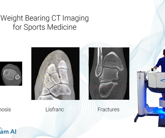
Weight Bearing CT Imaging for Sports Medicine

CurveBeam AI

Common Indications Syndesmosis Provide increased sensitivity and specificity over radiographs 1. Can Weight-Bearing Computed Tomography Be a Game-Changer in the Assessment of Ankle Sprain and Ankle Instability? Cone beam CT of the musculoskeletal system: clinical applications. 2018 Feb;9(1):35-45. Epub 2018 Jan 4.

article thumbnail

WBCT Indications Series: Midfoot Arthritis

CurveBeam AI

Diagnosis An accurate diagnosis and grading of the severity of osteoarthritic joints in the midfoot have been shown to be clinically relevant in treating the pathology early in its course and avoiding late-stage invasive procedures such as arthrodesis 3.​ Epub 2018 Oct 8. Images demonstrated severe degeneration of the midfoot joints.

Insiders

Sign Up for our Newsletter

This site is protected by reCAPTCHA and the Google Privacy Policy and Terms of Service apply.

Trending Sources

article thumbnail

WBCT Indication Series: End Stage Ankle Arthritis

CurveBeam AI

Radiographs demonstrated tibiotalar arthritis as well as adjacent-joint arthritis. His clinical interests include ankle arthritis, neuropathic disease, forefoot reconstruction, and deformity. (1) The Assessment of Ankle Osteoarthritis with Weight-Bearing Computed Tomography. Epub 2018 Oct 8. Foot Ankle Clin.

article thumbnail

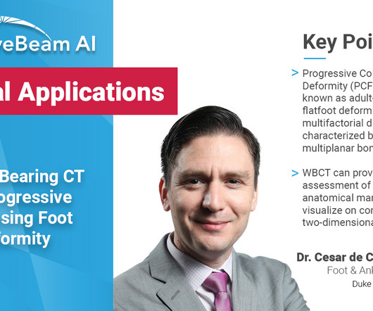
WBCT Indications Series: PCFD

CurveBeam AI

A weight bearing CT scan can: Provide an assessment of important anatomical markers of pronounced hindfoot deformity and peritalar subluxation (PTS), difficult to visualize on conventional two-dimensional radiographs 1. Epub 2018 Oct 8. Weightbearing Computed Tomography for Assessment of Foot and Ankle Deformities: The Iowa Experience.

article thumbnail

Lens Subluxation

Global Radiology CME

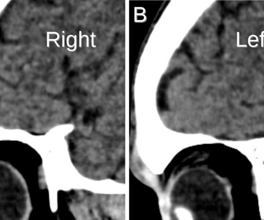
Computed tomography is also an alternative method for lens subluxation which again can show deviation of the lens (Figs. Radiographics. Sai is also a member of the Gold Humanism Honor Society and is involved with giving back to the community at a local free clinic as a medical assistant. 2006;10(4):345-350. J Emerg Med.

article thumbnail

A brief history of radiology

Radiology Cafe

Photoprint from radiograph by W.K. 3) In the early twentieth century, it was a common goal for investigators to try to find a way to separate the superimposed shadows that were recorded when a complex structure was shown on a radiograph. (3) 3) This is what is known as tomography. This is now known as ‘Hand mit Ringen’. (1)

article thumbnail

The ED Guide to Neuroimaging: Part 1

Northwestern EM

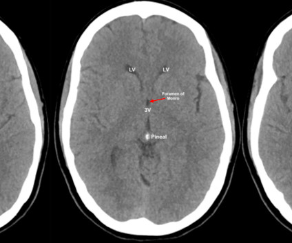
When evaluating a CT brain the following, certain cisterns have clinical relevance for potential herniation syndromes, layering of subarachnoid blood, and/or the significant structures that run through them. Computed Tomography. RadioGraphics, 1998; 18(1):151-163 3. In Clinical Neuroanatomy, 28th Edition (2013).