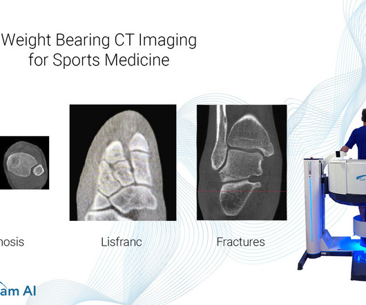
article thumbnail

WBCT Indications Series: Midfoot Arthritis

CurveBeam AI

In addition, WBCT better quantifies 3 the structural deformity of Chopart, talonavicular, and calcaneocuboid joints when compared to conventional radiography and non-weight bearing computed tomography images. However, the severity of deformity and involved joints were difficult to determine on plain radiographs alone.

article thumbnail

WBCT Indications Series: PCFD

CurveBeam AI

A weight bearing CT scan can: Provide an assessment of important anatomical markers of pronounced hindfoot deformity and peritalar subluxation (PTS), difficult to visualize on conventional two-dimensional radiographs 1. Epub 2018 Oct 8. Weightbearing Computed Tomography for Assessment of Foot and Ankle Deformities: The Iowa Experience.

Insiders

Sign Up for our Newsletter

This site is protected by reCAPTCHA and the Google Privacy Policy and Terms of Service apply.

Trending Sources

article thumbnail

WBCT Indication Series: End Stage Ankle Arthritis

CurveBeam AI

Radiographs demonstrated tibiotalar arthritis as well as adjacent-joint arthritis. The Assessment of Ankle Osteoarthritis with Weight-Bearing Computed Tomography. Characteristics of medial gutter arthritis on weightbearing CT and plain radiograph. Epub 2018 Oct 8. Foot Ankle Clin. 2022 Mar;27(1):13-36. 2021.11.001.

article thumbnail

Lens Subluxation

Global Radiology CME

Computed tomography is also an alternative method for lens subluxation which again can show deviation of the lens (Figs. Radiographics. Anterior dislocation of the lens is serious as it may lead to angle-closure glaucoma [3,4]. Lens subluxation can be diagnosed by ultrasound which shows deviation of the lens (Fig. J Emerg Med.

article thumbnail

A brief history of radiology

Radiology Cafe

Photoprint from radiograph by W.K. 3) In the early twentieth century, it was a common goal for investigators to try to find a way to separate the superimposed shadows that were recorded when a complex structure was shown on a radiograph. (3) 3) This is what is known as tomography. 2018 Aug; 18(8): 500–510. Röntgen, 1895.

article thumbnail

The ED Guide to Neuroimaging: Part 1

Northwestern EM

David Rusinak, MD Assistant Professor of Radiology, Northwestern Medicine How to cite this post [Peer-Reviewed, Web Publication] Whipple T, Gappmeier V (2018, April 23 ). Computed Tomography. RadioGraphics, 1998; 18(1):151-163 3. Demystifying the Hand Exam. Expert Commentary by Rusinak D ]. Shaw AS, Prokop M. McKetty MH.

article thumbnail

Emergency Guide to Stroke Neuroimaging

Northwestern EM

Last Update: September 14, 2018. Magnetic resonance imaging and computed tomography in emergency assessment of patients with suspected acute stroke: a prospective comparison. Contrast enhancement and contrast extravasation on computed tomography after intra-arterial thrombolysis in patients with acute ischemic stroke.