This site uses cookies to improve your experience. To help us insure we adhere to various privacy regulations, please select your country/region of residence. If you do not select a country, we will assume you are from the United States. Select your Cookie Settings or view our Privacy Policy and Terms of Use.
Cookie Settings
Cookies and similar technologies are used on this website for proper function of the website, for tracking performance analytics and for marketing purposes. We and some of our third-party providers may use cookie data for various purposes. Please review the cookie settings below and choose your preference.
Used for the proper function of the website
Used for monitoring website traffic and interactions
Cookie Settings
Cookies and similar technologies are used on this website for proper function of the website, for tracking performance analytics and for marketing purposes. We and some of our third-party providers may use cookie data for various purposes. Please review the cookie settings below and choose your preference.
Strictly Necessary: Used for the proper function of the website
Performance/Analytics: Used for monitoring website traffic and interactions
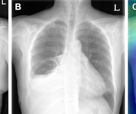
Radiologists routinely compare the current and previous chest radiographs during interpretation to enhance the sensitivity for change detection and provide information for differential diagnosis. Example of triage of no change in a pair of chest radiographs in the emergency department. (A) Image courtesy of Radiology.
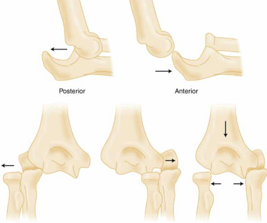
2018 Jun;54(6):849-854. Epub 2018 Apr 19. 2018 Apr 4;100(7):e46. Treasure Island (FL): StatPearls Publishing; 2024 Jan–. PMID: 30521261 Gottlieb M, Schiebout J. Elbow Dislocations in the Emergency Department: A Review of Reduction Techniques. J Emerg Med. doi: 10.1016/j.jemermed.2018.02.011. 2018.02.011. J Am Acad Orthop Surg.
The researchers included data from 1,242 patients recruited between 2013 and 2018 and divided them into training (n = 1,014) and testing groups (n = 228). They collected imaging data to establish deep-learning models using ResNet50. They used area under the receiver operating curve (AUC) to measure the predictive performance of the models.
To bridge this knowledge gap, the group analyzed 327 PET/CT diagnostic radiology reports between January 2001 and December 2018 at their institution that included descriptions of the finding. These signs often prompt otolaryngology referral to rule out malignancy yet the true risk based on the finding is unknown, they noted.
In 2018, Fishman received an endowed professorship from Johns Hopkins. I'm a radiographer,' " Stewart recalled. Throughout the 1980s, he worked on 3D imaging with Pixar Animation Studios and continues this work with firms such as Microsoft and Nvidia. I thought, 'I'm not a teacher. But Hennessy encouraged her.
In 2018 he received an endowed professorship from the university. Stewart is also co-author of the upcoming first edition of "Imaging Informatics: An Introduction" and the sole author of an Informatics in Medical Imaging chapter in the sixth edition of the textbook “Principles of Radiographic Imaging: An Art and Science."
And for the first time since 2018, physician burnout did not win the Minnies award for Biggest Threat to Radiology. Notably, AI was even more prevalent this year among the Minnies winners than in the past. The technology figured prominently in five Minnies categories, including Hottest Clinical Procedure.
mtaschetta-millane Thu, 07/25/2024 - 09:30 July 25, 2024 — The radiology gender gap is decreasing, but there remains work to be done, according to an editorial published today in RadioGraphics , a journal of the Radiological Society of North America ( RSNA ).
Radiographs demonstrated tibiotalar arthritis as well as adjacent-joint arthritis. Characteristics of medial gutter arthritis on weightbearing CT and plain radiograph. Epub 2018 Oct 8. Predict the likelihood of periprosthetic cyst formation based on varus/valgus 3. The calcaneal position was difficult to determine on X-Ray.
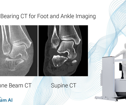
However, the severity of deformity and involved joints were difficult to determine on plain radiographs alone. As shown here, subluxation at the first and second TMT joints was better visualized on WBCT (bottom two scans) images than conventional radiographs (top scan). Epub 2018 Oct 8. PMID: 32851880. (2) Foot Ankle Int.
Kim Mason Kim Mason, an Audit and Research Radiographer for Mid Yorkshire Teaching Hospitals Trust, talks about their role as well as the value of radiographer engagement in research activities and how to get involved. Hi, I’m Kim and I am an alternative-styled, funky-haired, septum-pierced, disabled Audit and Research Radiographer.
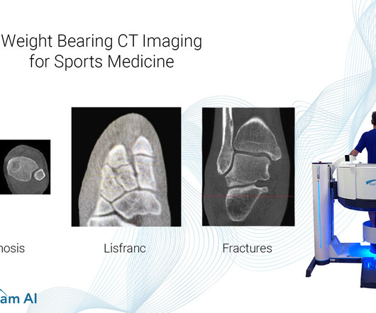
Common Indications Syndesmosis Provide increased sensitivity and specificity over radiographs 1. 2018 Feb;9(1):35-45. Epub 2018 Jan 4. Differentiate pathology from natural variability in patient anatomy via contralateral comparison to uninjured ankle as internal control 2. Help detect subtle syndesmosis injuries 1.
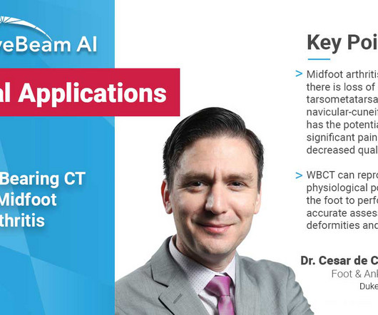
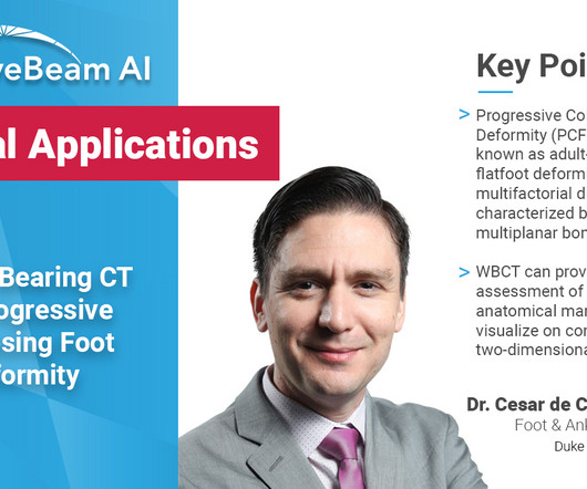
Common Indications Syndesmosis Provide increased sensitivity and specificity over radiographs 1. Flat Foot Provide an assessment of important anatomical markers of pronounced hindfoot deformity and peritalar subluxation (PTS), difficult to visualize on conventional two-dimensional radiographs 2. Epub 2018 Oct 8. Foot Ankle Int.
A weight bearing CT scan can: Provide an assessment of important anatomical markers of pronounced hindfoot deformity and peritalar subluxation (PTS), difficult to visualize on conventional two-dimensional radiographs 1. Epub 2018 Oct 8. PMID: 32851880. (2) 2) Jeng CL, Rutherford T, Hull MG, Cerrato RA, Campbell JT. Foot Ankle Int.
Epub 2018 Oct 6. Study subjects were stratified by the institution and by NIHSS score (≤15 or >15). Neurologic deficits were assessed at 72 hours, 1 month, and 3 months after randomization. PMID: 35963261 Sansevere AJ, Hahn CD, Abend NS. Conventional and quantitative EEG in status epilepticus. 2019 May;68:38-45. doi: 10.1016/j.seizure.2018.09.011.
Radiographics. Sai first realized his interest in radiology while he was conducting research in radiomics at the University of Michigan , where he graduated from in 2018 with a Bachelor of Science degree in Neuroscience. 2006;10(4):345-350. doi: 10.1016/j.jaapos.2006.01.218 2006.01.218 Kubal WS. Imaging of orbital trauma. J Emerg Med.
AI can process a massive number of medical images and automatically recognize radiographic characteristics. In a 2018 article , Jeffrey Golden, MD stated that AI can improve productivity by identifying features of interest in images before a human clinician reviews the data.
and to many people from the team at Addenbrooke’s, including a medical physicist and a research radiographer. Listen to “A Sense of Tumour” here About Angela Young Angela Young founded Cambridge Podcasts in 2018 to help clients showcase their expertise and establish themselves as the go-to person in their field.
David Rusinak, MD Assistant Professor of Radiology, Northwestern Medicine How to cite this post [Peer-Reviewed, Web Publication] Whipple T, Gappmeier V (2018, April 23 ). RadioGraphics, 1998; 18(1):151-163 3. A helpful tip is to look for subtle foci of intracranial air and soft tissue swelling which may direct you to a subtle fracture.
RadioGraphics. Paper presented at: MIT Technology Review EmTech Digital Conference; 2018. American College of Radiology. ACR Commission on Human Resources Workforce Survey. Harvey HB, et al. The Future of Radiology: Artificial Intelligence and Advanced Technologies. 2023;43(3):e230017. Society of Interventional Radiology. Hassabis D.
RadioGraphics. Paper presented at: MIT Technology Review EmTech Digital Conference; 2018. American College of Radiology. ACR Commission on Human Resources Workforce Survey. Harvey HB, et al. The Future of Radiology: Artificial Intelligence and Advanced Technologies. 2023;43(3):e230017. Society of Interventional Radiology. Hassabis D.
Avery’s Diseases of the Newborn, Elsevier, 2018, pp. Obstetric Imaging: Fetal Diagnosis and Care, Elsevier, 2018, pp. Seeing the radiographic images made medical education come to life for him. National Institute of Neurological Disorders and Stroke. link] Huang SB, Doherty D. doi: 10.1016/B978-0-323-40139-5.00059-0.
JACC Cardiovasc Interv 2018. in the paper but 2.7% to ≈0.99 (p<0.001) Mean MPI/Tei Index≈ 0.47 A Randomized Trial of the Optimum Duration of Acoustic Pulse Thrombolysis Procedure in Acute Intermediate-Reisk Pulmonary Embolism: The OPTALYSE PE Trial. PMID: 30025734 Sharifi M et al. Am J Cardiol 2013. PMID: 23102885 Aykan AC et al.
Photoprint from radiograph by W.K. 3) In the early twentieth century, it was a common goal for investigators to try to find a way to separate the superimposed shadows that were recorded when a complex structure was shown on a radiograph. (3) 2018 Aug; 18(8): 500–510. This is now known as ‘Hand mit Ringen’. (1) Röntgen, 1895.
In this study, the researchers tested the model in a cohort of 36,924 asymptomatic Asian individuals (mean chronological age, 58 years old ± 7 years; 22,352 male and 14,572 female) who underwent imaging as part of regular health check-ups at their hospitals between January 2004 and June 2018.
Last Update: September 14, 2018. Last Updated: 2018. Radiographics. Effect of treatment delay, age, and stroke severity on the effects of intravenous thrombolysis with alteplase for acute ischaemic stroke: a meta-analysis of individual patient data from randomised trials. 2014 Nov 29;384(9958):1929-35. Filho JO, Samuels OB.
Of the patients with radiographically proven adverse radiation effects (AREs; 15%), 4 were symptomatic. J Neurosurg 2022;137(August):533–43 This single-center study included 358 eligible patients with histologically confirmed isocitrate dehydrogenase (IDH)–wild-type GBM from November 1, 2005, to December 31, 2018.
We organize all of the trending information in your field so you don't have to. Join 5,000 users and stay up to date on the latest articles your peers are reading.
You know about us, now we want to get to know you!
Let's personalize your content
Let's get even more personalized
We recognize your account from another site in our network, please click 'Send Email' below to continue with verifying your account and setting a password.


Let's personalize your content