Remove 2019 Remove Clinic Remove Radiographer Remove Radiography
article thumbnail

Opportunity for osteoporosis check on lumbar spine plain radiographs

AuntMinnie

. | W3-SSMK08-4 | Room E450A A deep learning-based framework for automated screening of osteoporosis on lumbar spine plain radiographs shows potential as another way to opportunistically make use of imaging studies performed for other indications, according to this presentation.

article thumbnail

Meet the Minnies 2024 semifinal candidates

AuntMinnie

Assessment of Claimant, Clinical, and Financial Characteristics of Teleradiology Medical Malpractice Cases. Commercially Available Chest Radiograph AI Tools for Detecting Airspace Disease, Pneumothorax, and Pleural Effusion. Generative Artificial Intelligence for Chest Radiograph Interpretation in the Emergency Department.

Insiders

Sign Up for our Newsletter

This site is protected by reCAPTCHA and the Google Privacy Policy and Terms of Service apply.

Trending Sources

article thumbnail

Open Disclosure in Radiology

Everything Rad

By Professor Louise Rainford, Associate Dean of Radiography, UCD Dublin. How are radiographers treated when they admit to making a mistake? And are clinical staff treated the same across disciplines when they make mistakes? Two, periodically review with radiographers the process for reporting safety incidents.

article thumbnail

WBCT Indications Series: Midfoot Arthritis

CurveBeam AI

Diagnosis An accurate diagnosis and grading of the severity of osteoarthritic joints in the midfoot have been shown to be clinically relevant in treating the pathology early in its course and avoiding late-stage invasive procedures such as arthrodesis 3.​ 2019 Feb;40(2):152-158. PMID: 32851880.​ (2) Foot Ankle Int. Epub 2018 Oct 8.

article thumbnail

WBCT Indications Series: PCFD

CurveBeam AI

A weight bearing CT scan can: Provide an assessment of important anatomical markers of pronounced hindfoot deformity and peritalar subluxation (PTS), difficult to visualize on conventional two-dimensional radiographs 1. 2019 Feb;40(2):152-158. PMID: 32851880.​ (2) 2) Jeng CL, Rutherford T, Hull MG, Cerrato RA, Campbell JT. 2020.109419.

article thumbnail

Lisfranc Fracture Dislocation

Global Radiology CME

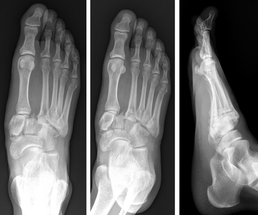
A) AP radiograph of Lisfranc Fracture Dislocation demonstrates the circled “fleck sign” or Lisfranc ligament avulsion fracture fragment. (B) C) The lateral radiograph notes with a circle, the dorsal sub dislocation of the metatarsal base. Radiographs should be repeated after two weeks to ensure surgery is unnecessary.

article thumbnail

Minnies winners for 2023 highlight AI, innovation in radiology

AuntMinnie

The technology figured prominently in five Minnies categories, including Hottest Clinical Procedure. Pickhardt believes using additional clinical data on CT imaging could save healthcare costs, especially when AI tools are used in parallel, suggesting it is a cost-effective or even cost-saving strategy for personalized or precision medicine.

Radiology 132