Remove 2019 Remove Computed Tomography Remove Radiograph Remove Radiography
article thumbnail

WBCT Indications Series: Midfoot Arthritis

CurveBeam AI

In addition, WBCT better quantifies 3 the structural deformity of Chopart, talonavicular, and calcaneocuboid joints when compared to conventional radiography and non-weight bearing computed tomography images. However, the severity of deformity and involved joints were difficult to determine on plain radiographs alone.


article thumbnail

WBCT Indications Series: PCFD

CurveBeam AI

A weight bearing CT scan can: Provide an assessment of important anatomical markers of pronounced hindfoot deformity and peritalar subluxation (PTS), difficult to visualize on conventional two-dimensional radiographs 1. 2019 Feb;40(2):152-158. PMID: 32851880.​ (2) 2) Jeng CL, Rutherford T, Hull MG, Cerrato RA, Campbell JT. Leardini, A.,

article thumbnail

Lisfranc Fracture Dislocation

Global Radiology CME

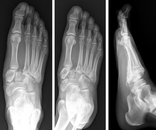
A) AP radiograph of Lisfranc Fracture Dislocation demonstrates the circled “fleck sign” or Lisfranc ligament avulsion fracture fragment. (B) C) The lateral radiograph notes with a circle, the dorsal sub dislocation of the metatarsal base. Radiographs should be repeated after two weeks to ensure surgery is unnecessary.