article thumbnail

Opportunity for osteoporosis check on lumbar spine plain radiographs

AuntMinnie

. | W3-SSMK08-4 | Room E450A A deep learning-based framework for automated screening of osteoporosis on lumbar spine plain radiographs shows potential as another way to opportunistically make use of imaging studies performed for other indications, according to this presentation.

article thumbnail

Open Disclosure in Radiology

Everything Rad

By Professor Louise Rainford, Associate Dean of Radiography, UCD Dublin. How are radiographers treated when they admit to making a mistake? A patient might slip and fall in the exam room; a radiographer might inadvertently touch or “catch” the patient with equipment. Are the processes for reporting well known?

Insiders

Sign Up for our Newsletter

This site is protected by reCAPTCHA and the Google Privacy Policy and Terms of Service apply.

Trending Sources

article thumbnail

WBCT Indications Series: Midfoot Arthritis

CurveBeam AI

In addition, WBCT better quantifies 3 the structural deformity of Chopart, talonavicular, and calcaneocuboid joints when compared to conventional radiography and non-weight bearing computed tomography images. However, the severity of deformity and involved joints were difficult to determine on plain radiographs alone. Foot Ankle Int.

article thumbnail

WBCT Indications Series: PCFD

CurveBeam AI

A weight bearing CT scan can: Provide an assessment of important anatomical markers of pronounced hindfoot deformity and peritalar subluxation (PTS), difficult to visualize on conventional two-dimensional radiographs 1. 2019 Feb;40(2):152-158. PMID: 32851880.​ (2) 2) Jeng CL, Rutherford T, Hull MG, Cerrato RA, Campbell JT. 2020.109419.

article thumbnail

Lisfranc Fracture Dislocation

Global Radiology CME

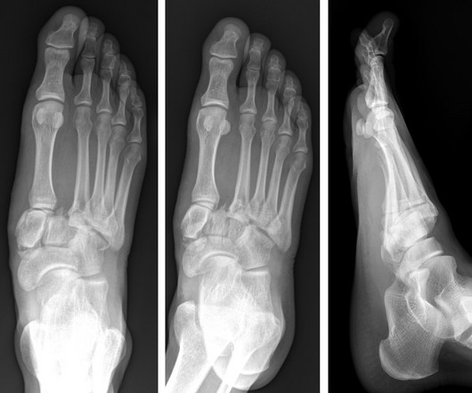
A) AP radiograph of Lisfranc Fracture Dislocation demonstrates the circled “fleck sign” or Lisfranc ligament avulsion fracture fragment. (B) C) The lateral radiograph notes with a circle, the dorsal sub dislocation of the metatarsal base. Radiographs should be repeated after two weeks to ensure surgery is unnecessary.

article thumbnail

A podcast radiating positivity

The British Institute of Radiology

I had been having regular follow up scans, and in 2019, it was thought the growth was significant enough to consider radiotherapy. and to many people from the team at Addenbrooke’s, including a medical physicist and a research radiographer.

article thumbnail

Minnies winners for 2023 highlight AI, innovation in radiology

AuntMinnie

The first female editor in chief of RSNA’s peer-reviewed RadioGraphics journal since 2021, Menias splits her time between Mayo Clinic, where she’s a specialist in abdominal imaging, and the journal, where she is achieving her first goals of expanding RadioGraphics as a learning platform.

Radiology 132