Remove 2021 Remove Clinic Remove Computed Tomography Remove Radiography
article thumbnail

WBCT Indications Series: Midfoot Arthritis

CurveBeam AI

Diagnosis An accurate diagnosis and grading of the severity of osteoarthritic joints in the midfoot have been shown to be clinically relevant in treating the pathology early in its course and avoiding late-stage invasive procedures such as arthrodesis 3.​ 2021 Jun;42(6):757-767. Epub 2021 Jan 27. Sci Rep 11, 16139 (2021).

article thumbnail

WBCT Indications Series: PCFD

CurveBeam AI

In addition, WBCT better quantifies the structural deformity of PCFD compared to conventional radiography and non-weight bearing CT images 1 ​​. 2021 Jun;42(6):757-767. Epub 2021 Jan 27. Weightbearing Computed Tomography for Assessment of Foot and Ankle Deformities: The Iowa Experience. Sci Rep 11, 16139 (2021).

Insiders

Sign Up for our Newsletter

This site is protected by reCAPTCHA and the Google Privacy Policy and Terms of Service apply.

Trending Sources

article thumbnail

WBCT Indications Series: Lisfranc

CurveBeam AI

2021, May 20). MedShape and Curvebeam announce initiation of joint prospective clinical study. 2021 Nov;39(11):2497-2505. Epub 2021 Jan 6. Comparative assessment of midfoot osteoarthritis diagnostic sensitivity using weightbearing computed tomography vs weightbearing plain radiography. CurveBeam AI.

article thumbnail

WBCT Indication Series: Hallux Valgus

CurveBeam AI

Comparative assessment of midfoot osteoarthritis diagnostic sensitivity using weightbearing computed tomography vs weightbearing plain radiography. 2021 Jan;134:109419. Journal of the Foot & Ankle. 10.30795/jfootankle.2022.v16.1674. v16.1674. (4) 4) Steadman J, Sripanich Y, Rungprai C, Mills MK, Saltzman CL, Barg A.

article thumbnail

Lisfranc Fracture Dislocation

Global Radiology CME

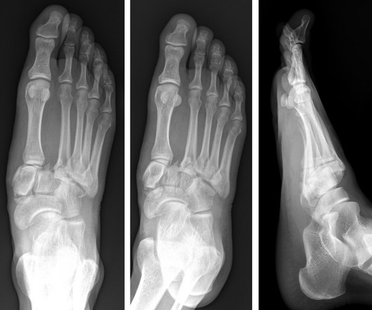
A computed tomography scan will better assist with diagnosis and help with planning if surgery is necessary [3]. Is the diagnostic validity of conventional radiography for Lisfranc injury acceptable? In this case the lateral view shows a dorsal sub dislocation of the metatarsal base (Fig. J Foot Ankle Res. 2023;16(1):9.