Remove 2021 Remove Computed Tomography Remove Imaging Remove Radiography
article thumbnail

WBCT Indications Series: Midfoot Arthritis

CurveBeam AI

In addition, WBCT better quantifies 3 the structural deformity of Chopart, talonavicular, and calcaneocuboid joints when compared to conventional radiography and non-weight bearing computed tomography images. Images demonstrated severe degeneration of the midfoot joints. 2021 Jun;42(6):757-767. Eur J Radiol.


article thumbnail

WBCT Indications Series: PCFD

CurveBeam AI

In addition, WBCT better quantifies the structural deformity of PCFD compared to conventional radiography and non-weight bearing CT images 1 ​​. 2021 Jun;42(6):757-767. Epub 2021 Jan 27. Weightbearing Computed Tomography for Assessment of Foot and Ankle Deformities: The Iowa Experience. 2021 Jan;134:109419.

Insiders

Sign Up for our Newsletter

This site is protected by reCAPTCHA and the Google Privacy Policy and Terms of Service apply.

Trending Sources

article thumbnail

WBCT Indication Series: Hallux Valgus

CurveBeam AI

Treatment Planning Because hallux valgus is a three-dimensional deformity, a three-dimensional, weight bearing imaging modality can assist in planning surgical indications and correction.Surgeons can better determine where along the medial column the deformity should be addressed. 2021 Jan;134:109419. Journal of the Foot & Ankle.

article thumbnail

Lisfranc Fracture Dislocation

Global Radiology CME

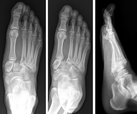
Imaging and Case Analysis: Radiographic images demonstrate misalignment of the medial side of the second metatarsal with the medial side of the middle cuneiform bone, as seen in this case. A computed tomography scan will better assist with diagnosis and help with planning if surgery is necessary [3]. J Foot Ankle Res.