Remove 2021 Remove Computed Tomography Remove Radiograph
article thumbnail

WBCT Indications Series: Midfoot Arthritis

CurveBeam AI

In addition, WBCT better quantifies 3 the structural deformity of Chopart, talonavicular, and calcaneocuboid joints when compared to conventional radiography and non-weight bearing computed tomography images. However, the severity of deformity and involved joints were difficult to determine on plain radiographs alone.

article thumbnail

WBCT Indications Series: Lisfranc

CurveBeam AI

Diagnosis The diagnosis of Lisfranc injuries may be challenging on plain radiographs alone. Radiographs were indeterminate. 2021, May 20). 2021 Nov;39(11):2497-2505. Epub 2021 Jan 6. 2021 Jan;134:109419. A WBCT scan may also provide additional information about concomitant injuries and fractures.​
Insiders

Sign Up for our Newsletter

This site is protected by reCAPTCHA and the Google Privacy Policy and Terms of Service apply.

Trending Sources

article thumbnail

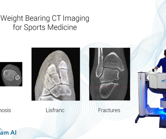
Weight Bearing CT Imaging for Sports Medicine

CurveBeam AI

Common Indications Syndesmosis Provide increased sensitivity and specificity over radiographs 1. Can Weight-Bearing Computed Tomography Be a Game-Changer in the Assessment of Ankle Sprain and Ankle Instability? 2021 Nov;39(11):2497-2505. Epub 2021 Jan 6. Help detect subtle syndesmosis injuries 1. Foot Ankle Clin.

article thumbnail

WBCT Indications Series: PCFD

CurveBeam AI

A weight bearing CT scan can: Provide an assessment of important anatomical markers of pronounced hindfoot deformity and peritalar subluxation (PTS), difficult to visualize on conventional two-dimensional radiographs 1. 2021 Jun;42(6):757-767. Epub 2021 Jan 27. Sci Rep 11, 16139 (2021). 2021 Jan;134:109419.

article thumbnail

WBCT Indication Series: End Stage Ankle Arthritis

CurveBeam AI

Radiographs demonstrated tibiotalar arthritis as well as adjacent-joint arthritis. The Assessment of Ankle Osteoarthritis with Weight-Bearing Computed Tomography. 2021 Feb;42(2):200214. Characteristics of medial gutter arthritis on weightbearing CT and plain radiograph. 2021 Aug;50(8):1575-1583. 2021.11.001.

article thumbnail

FDA approves Bayer’s Ultravist (iopromide) Injection for Contrast-enhanced Mammography

Imaging Technology

In 2019, the MEDRAD Stellant FLEX Computed Tomography ( CT ) Injection System with Certegra Workstation was also cleared in the U.S. RadioGraphics 2019 39:7, 1907-1920. RadioGraphics 2021 41:3, 829-839. Updated March 26, 2021. for use in CEM. Breast Density on a Mammogram. Updated April 4, 2023.

article thumbnail

Lisfranc Fracture Dislocation

Global Radiology CME

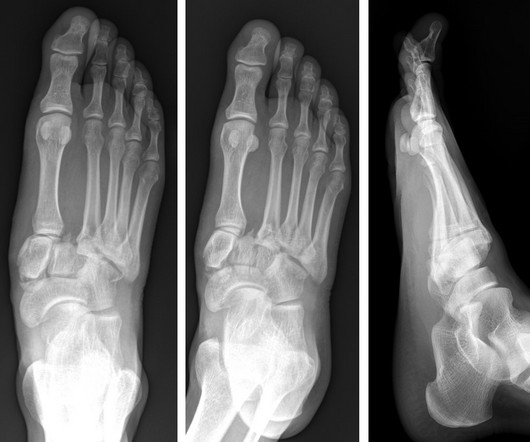
A) AP radiograph of Lisfranc Fracture Dislocation demonstrates the circled “fleck sign” or Lisfranc ligament avulsion fracture fragment. (B) C) The lateral radiograph notes with a circle, the dorsal sub dislocation of the metatarsal base. Radiographs should be repeated after two weeks to ensure surgery is unnecessary.