Remove 2021 Remove Hospital Remove Radiographer Remove Radiologist
article thumbnail

Meet the Minnies 2024 semifinal candidates

AuntMinnie

tesla MRI AI body composition analysis Cardiac PET Cryo/thermoablation CT colonography Genicular artery embolization Hyperpolarized xenon-129 MRI PET/MRI Photon-counting CT Radiomics Theranostics Whole-body MRI screening Image of the Year 3D PET/MR image.


article thumbnail

Responding to a Ransomware Attack in Radiology

Everything Rad

Reading Time: 8 minutes read By Dr. Niall Sheehy, MB, MRCP, FFR RCSI, St James’s Hospital -Trinity College Dublin. In May 2021, St James’s Hospital in Dublin was one of 54 public hospitals affected when the Health Service Executive (HSE) was the victim of a Cyber Attack by Conti, a sophisticated, financially-motivated criminal gang.

PACS 98
Insiders

Sign Up for our Newsletter

This site is protected by reCAPTCHA and the Google Privacy Policy and Terms of Service apply.

Trending Sources

article thumbnail

Lisfranc Fracture Dislocation

Global Radiology CME

A) AP radiograph of Lisfranc Fracture Dislocation demonstrates the circled “fleck sign” or Lisfranc ligament avulsion fracture fragment. (B) C) The lateral radiograph notes with a circle, the dorsal sub dislocation of the metatarsal base. Radiographs should be repeated after two weeks to ensure surgery is unnecessary.

article thumbnail

Type 3 Cuboid Fracture

Global Radiology CME

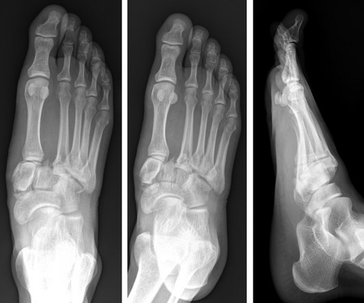
A) Dorsoplantar radiograph of the foot demonstrating an isolated fracture of the cuboid with possible extension into the tarsometatarsal joint. (B) B) Medial oblique radiograph of the foot demonstrating an isolated fracture of the cuboid. Radiographic evidence can support the diagnosis. Trauma in a 8 year old female.

article thumbnail

Capsule Retention Following Capsule Endoscopy

Global Radiology CME

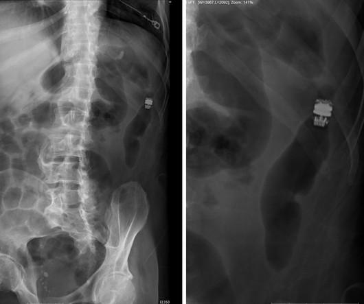
Frontal abdomen radiograph demonstrates foreign body consistent with capsule endoscopy device (pill cam) in descending colon. A prospective study of the utility of abdominal radiographs after capsule endoscopy for the diagnosis of capsule retention. 61-year-old male with abdominal pain 15 days after capsule endoscopy. doi: 10.3748/wjg.15.2401

X-ray 52
article thumbnail

Emphysematous Pyelonephritis

Global Radiology CME

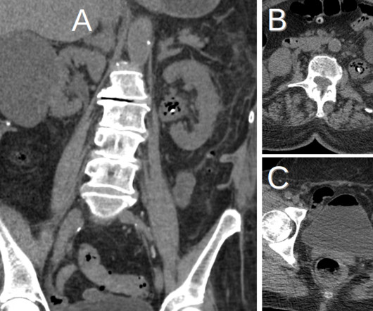
KUB indicates kidneys, ureter, and bladder (plain abdominal radiograph); CT, computed tomography; and PCD, percutaneous catheter drainage. Date of download: 7/25/2021 EPN is a life-threatening condition that warrants an immediate treatment. RadioGraphics. 2000;160(6):797-805. doi:10.1001/archinte.160.6.797 2000;160(6):797-805.

article thumbnail

Nanox.ARC Imaging System Receives FDA Clearance, Pioneering a New Era in Medical Imaging

Imaging Technology

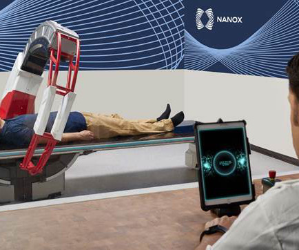
The Company received 510(k) clearance to market its single-source X-ray device, known as the Nanox Cart X-Ray System, in April 2021. The multi-source Nanox.ARC has the ability to reconstruct a series of 2D projection images into a stack of tomograms (or slices) of the imaged object, forming a 3D visualization.