Remove 2021 Remove Magnetic Resonance Imaging Remove Radiograph
article thumbnail

Meet the Minnies 2024 semifinal candidates

AuntMinnie

Image from Fangyi Liu, MD, of the Chinese PLA General Hospital, et al. Scientific Paper of the Year Advanced Practice Provider Procedures Commonly Performed in Interventional Radiology: Medicare Volume Trends From 2010 to 2021. Generative Artificial Intelligence for Chest Radiograph Interpretation in the Emergency Department.

article thumbnail

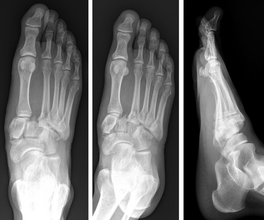
Lisfranc Fracture Dislocation

Global Radiology CME

A) AP radiograph of Lisfranc Fracture Dislocation demonstrates the circled “fleck sign” or Lisfranc ligament avulsion fracture fragment. (B) C) The lateral radiograph notes with a circle, the dorsal sub dislocation of the metatarsal base. Radiographs should be repeated after two weeks to ensure surgery is unnecessary.

Insiders

Sign Up for our Newsletter

This site is protected by reCAPTCHA and the Google Privacy Policy and Terms of Service apply.

Trending Sources

article thumbnail

Type 3 Cuboid Fracture

Global Radiology CME

A) Dorsoplantar radiograph of the foot demonstrating an isolated fracture of the cuboid with possible extension into the tarsometatarsal joint. (B) B) Medial oblique radiograph of the foot demonstrating an isolated fracture of the cuboid. Radiographic evidence can support the diagnosis. Trauma in a 8 year old female.

article thumbnail

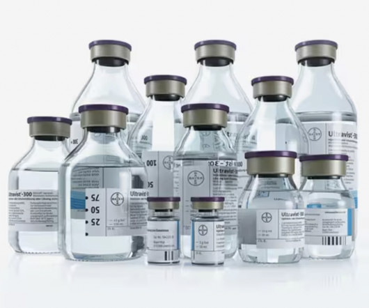
FDA approves Bayer’s Ultravist (iopromide) Injection for Contrast-enhanced Mammography

Imaging Technology

The approval expands upon Bayer's focus on breast imaging, with a portfolio that also includes Gadavist (gadobutrol) injection, a gadolinium-based contrast agent approved for use with MRI ( Magnetic Resonance Imaging ) to assess the presence and extent of malignant breast disease in adult patients. for use in CEM.

article thumbnail

Journal Scan – This Month in Other Journals, June 2023

AJNR

It has well-defined radiographic features and various clinical presentations. They described six radiographic phenotypes of CSVD: (1) recent small subcortical infarct, (2) white matter hyperintensity, (3) lacune of presumed vascular origin, (4) widened perivascular spaces, (5) cerebral microbleed, and (6) brain atrophy.

Disease 40