article thumbnail

Can We Upskill Radiographers through Artificial Intelligence? 

The British Institute of Radiology

Shamie Kumar describes how AI fits into a radiology clinical workflow and her perspective on how a clinical radiographer could use this to learn from and enhance their skills. If the AI findings are seen in PACS, how many radiographers actually log into PACS after taking a scan or X-ray? Can Radiographers Up-Skill?

article thumbnail

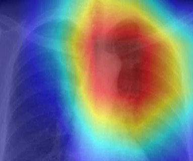
Deep-Learning Chest Radiograph Model Predicts Mortality for Community-Acquired Pneumonia

Imaging Technology

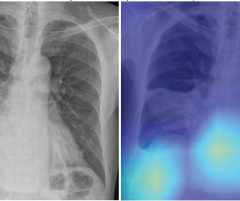
In this AJR accepted manuscript , a DL model was developed in 7,105 patients via one institution from March 2013 to December 2019 (3:1:1 allocation to training, validation, and internal test sets) to predict risk of all-cause mortality within 30 days after CAP diagnosis using patients’ initial chest radiograph. CURB-65 score). Hwang et al.

Insiders

Sign Up for our Newsletter

This site is protected by reCAPTCHA and the Google Privacy Policy and Terms of Service apply.

Trending Sources

article thumbnail

WBCT Indications Series: Lisfranc

CurveBeam AI

Diagnosis The diagnosis of Lisfranc injuries may be challenging on plain radiographs alone. Radiographs were indeterminate. 2021, May 20). 2021 Nov;39(11):2497-2505. Epub 2021 Jan 6. 2021 Jan;134:109419. A WBCT scan may also provide additional information about concomitant injuries and fractures.​

article thumbnail

WBCT Indications Series: Midfoot Arthritis

CurveBeam AI

However, the severity of deformity and involved joints were difficult to determine on plain radiographs alone. As shown here, subluxation at the first and second TMT joints was better visualized on WBCT (bottom two scans) images than conventional radiographs (top scan). 2021 Jun;42(6):757-767. Epub 2021 Jan 27.

article thumbnail

WBCT Indication Series: End Stage Ankle Arthritis

CurveBeam AI

Radiographs demonstrated tibiotalar arthritis as well as adjacent-joint arthritis. 2021 Feb;42(2):200214. Characteristics of medial gutter arthritis on weightbearing CT and plain radiograph. 2021 Aug;50(8):1575-1583. Epub 2021 Jan 7. Predict the likelihood of periprosthetic cyst formation based on varus/valgus 3​.

article thumbnail

Radiologists Must Monitor Novel Alzheimer’s Treatment Side Effect

Imaging Technology

In June 2021, the U.S. Most patients with asymptomatic ARIA meeting specific radiographic and clinical criteria may continue to receive treatment. The main pathologic feature of Alzheimer disease is a buildup of toxic amyloid-B. Disease-modifying drugs like monoclonal antibodies work by clearing toxic amyloid-B protein from the brain.

article thumbnail

Lisfranc Fracture Dislocation

Global Radiology CME

A) AP radiograph of Lisfranc Fracture Dislocation demonstrates the circled “fleck sign” or Lisfranc ligament avulsion fracture fragment. (B) C) The lateral radiograph notes with a circle, the dorsal sub dislocation of the metatarsal base. Radiographs should be repeated after two weeks to ensure surgery is unnecessary.