article thumbnail

Can We Upskill Radiographers through Artificial Intelligence? 

The British Institute of Radiology

Shamie Kumar describes how AI fits into a radiology clinical workflow and her perspective on how a clinical radiographer could use this to learn from and enhance their skills. If the AI findings are seen in PACS, how many radiographers actually log into PACS after taking a scan or X-ray? Can Radiographers Up-Skill?

article thumbnail

WBCT Indications Series: Lisfranc

CurveBeam AI

Diagnosis The diagnosis of Lisfranc injuries may be challenging on plain radiographs alone. Radiographs were indeterminate. 2021, May 20). 2021 Nov;39(11):2497-2505. Epub 2021 Jan 6. 2021 Jan;134:109419. A WBCT scan may also provide additional information about concomitant injuries and fractures.​

Insiders

Sign Up for our Newsletter

This site is protected by reCAPTCHA and the Google Privacy Policy and Terms of Service apply.

Trending Sources

article thumbnail

WBCT Indications Series: Midfoot Arthritis

CurveBeam AI

In addition, WBCT better quantifies 3 the structural deformity of Chopart, talonavicular, and calcaneocuboid joints when compared to conventional radiography and non-weight bearing computed tomography images. However, the severity of deformity and involved joints were difficult to determine on plain radiographs alone. Epub 2021 Jan 27.

article thumbnail

WBCT Indications Series: PCFD

CurveBeam AI

A weight bearing CT scan can: Provide an assessment of important anatomical markers of pronounced hindfoot deformity and peritalar subluxation (PTS), difficult to visualize on conventional two-dimensional radiographs 1. 2021 Jun;42(6):757-767. Epub 2021 Jan 27. Sci Rep 11, 16139 (2021). 2021 Jan;134:109419.

article thumbnail

WBCT and its Evolving Role in OA Research and Clinical Practice

CurveBeam AI

Key Points: Weight bearing CT (WBCT) can detect signs of osteoarthritis (OA), such as osteophytes, subchondral cysts, and joint space narrowing better than radiographs. Emerging evidence suggests WBCT reveals meniscal extrusion not detected by MRI and WBCT arthrography visualizes meniscal tears. Segal et al.

Clinic 52
article thumbnail

WBCT Indication Series: Hallux Valgus

CurveBeam AI

Diagnosis While radiographs are typically sufficient to make the diagnosis, WBCT scans may be useful to plan surgical treatment. Accurately assess sesamoid position as plain radiographs cannot determine whether the sesamoids have been reduced within their grooves 5. 2021 Jan;134:109419. Journal of the Foot & Ankle.

article thumbnail

Lisfranc Fracture Dislocation

Global Radiology CME

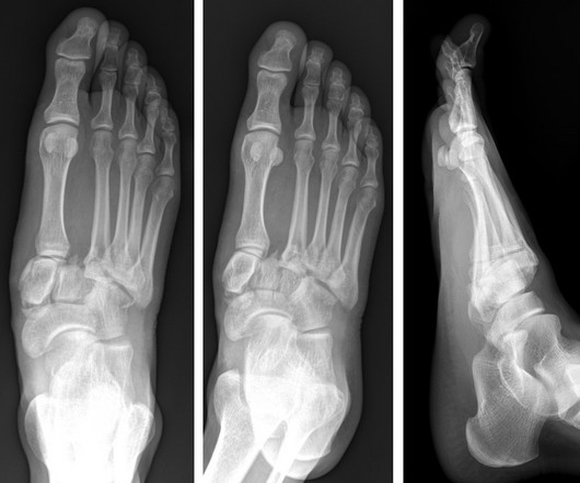
A) AP radiograph of Lisfranc Fracture Dislocation demonstrates the circled “fleck sign” or Lisfranc ligament avulsion fracture fragment. (B) C) The lateral radiograph notes with a circle, the dorsal sub dislocation of the metatarsal base. Radiographs should be repeated after two weeks to ensure surgery is unnecessary.