Remove 2022 Remove Computed Tomography Remove Diagnostic Remove Radiographer
article thumbnail

ECR: 3 tips for developing a successful cardiac imaging practice

AuntMinnie

"Together with our radiographers, I learned to scan cardiac patients and learned special anatomy from pediatric cardiologists and pediatric cardiac surgeons." He noted that in 2019, the European Society of Cardiology issued updated guidelines for diagnostic imaging of coronary artery disease (CAD), recommending noninvasive imaging (i.e.,

Imaging 104
article thumbnail

WBCT Indications Series: Lisfranc

CurveBeam AI

Diagnosis The diagnosis of Lisfranc injuries may be challenging on plain radiographs alone. Radiographs were indeterminate. 2022, February 24). Comparative assessment of midfoot osteoarthritis diagnostic sensitivity using weightbearing computed tomography vs weightbearing plain radiography. Foot Ankle Int.

Insiders

Sign Up for our Newsletter

This site is protected by reCAPTCHA and the Google Privacy Policy and Terms of Service apply.

Trending Sources

article thumbnail

Lisfranc Fracture Dislocation

Global Radiology CME

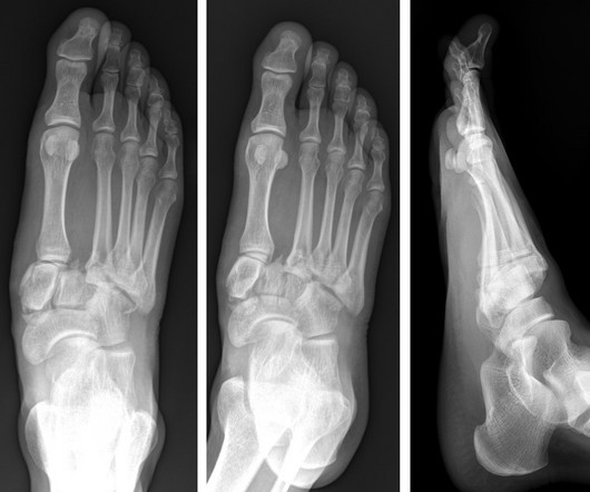
A) AP radiograph of Lisfranc Fracture Dislocation demonstrates the circled “fleck sign” or Lisfranc ligament avulsion fracture fragment. (B) C) The lateral radiograph notes with a circle, the dorsal sub dislocation of the metatarsal base. Radiographs should be repeated after two weeks to ensure surgery is unnecessary.

article thumbnail

WBCT Indication Series: Hallux Valgus

CurveBeam AI

Diagnosis While radiographs are typically sufficient to make the diagnosis, WBCT scans may be useful to plan surgical treatment. Accurately assess sesamoid position as plain radiographs cannot determine whether the sesamoids have been reduced within their grooves 5. FAI 2022. (2) Accurately assess healing in the 1st TMT joint 4.

article thumbnail

WBCT Indications Series: Syndesmosis

CurveBeam AI

A weight bearing CT scan can: Provide increased sensitivity and specificity over radiographs​. X-rays were non-diagnostic. Syndesmosis A syndesmotic ankle sprain, also known as high ankle sprain, is an injury to one or more of the ligaments comprising the distal tibiofibular syndesmosis.

X-ray 98
article thumbnail

Journal Scan – This Month in Other Journals, June 2023

AJNR

It has well-defined radiographic features and various clinical presentations. They described six radiographic phenotypes of CSVD: (1) recent small subcortical infarct, (2) white matter hyperintensity, (3) lacune of presumed vascular origin, (4) widened perivascular spaces, (5) cerebral microbleed, and (6) brain atrophy. 3 figures 4.

Disease 40