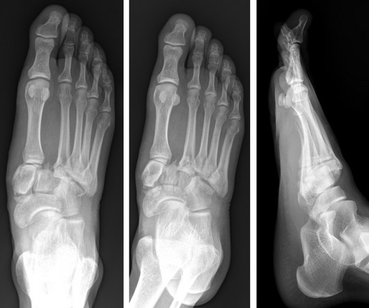
Lisfranc Fracture Dislocation
Global Radiology CME
NOVEMBER 10, 2023
Imaging and Case Analysis: Radiographic images demonstrate misalignment of the medial side of the second metatarsal with the medial side of the middle cuneiform bone, as seen in this case. A computed tomography scan will better assist with diagnosis and help with planning if surgery is necessary [3]. Lisfranc Dislocation.


Let's personalize your content