This site uses cookies to improve your experience. To help us insure we adhere to various privacy regulations, please select your country/region of residence. If you do not select a country, we will assume you are from the United States. Select your Cookie Settings or view our Privacy Policy and Terms of Use.
Cookie Settings
Cookies and similar technologies are used on this website for proper function of the website, for tracking performance analytics and for marketing purposes. We and some of our third-party providers may use cookie data for various purposes. Please review the cookie settings below and choose your preference.
Used for the proper function of the website
Used for monitoring website traffic and interactions
Cookie Settings
Cookies and similar technologies are used on this website for proper function of the website, for tracking performance analytics and for marketing purposes. We and some of our third-party providers may use cookie data for various purposes. Please review the cookie settings below and choose your preference.
Strictly Necessary: Used for the proper function of the website
Performance/Analytics: Used for monitoring website traffic and interactions
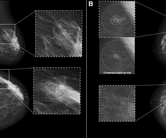
This study] suggests a differential reliance on decision support related to whether that originated from AI CAD or from a fellow radiologist, the authors wrote in an article published March 18 in Radiology. All images and caption courtesy of the RSNA. The full article and accompanying commentary can be found here and here.
FDG-PET imaging shows promise for use as a diagnostic criterion for neurosarcoidosis, with a recent case series illustrating the approach was effective when gold-standard approaches were not, according to a group of researchers in Berlin. “To The article was published January 25 in Neurological Research and Practice.
Technetium-99m (Tc-99m) methyl diphosphonate (MDP) bone scans are a potentially viable noninvasive option for diagnosing calciphylaxis, according to a team at the University of Massachusetts in Worcester, MA. and Canada, which reported the sensitivity of bone scans for diagnosing calciphylaxis to be 62.5%, 89%, and 94.4%.
Lymphoscintigraphy (LSG) is recommended but seldom used to diagnose lymphedema in real-world settings in the U.S., and 4.4%), while these imaging studies were less frequently used in the upper extremity, around 1%. The full article is available here.
PET/MRI imaging shows promise in diagnosing fevers or inflammation of unknown origin and may have advantages over PET/CT, according to a study published January 3 in the European Journal of Radiology. T2 IDEAL WATER image (B) shows increased T2 signal; LAVA WATER (C) image shows mild enhancement in these muscles.
F-18 FAPI-PET/CT is superior to F-18 FDG-PET/CT for diagnosing and staging patients with pancreatic cancer, according to a study published January 4 in the Journal of Nuclear Medicine. Typical PET (top), PET/CT (middle), and CT and MR (bottom) images of primary tumor obtained using both radiotracers in representative patients (A and B).
We hope you enjoy our article as well as the opportunity to hear directly from the recipients. His lab team is currently focused on "implementing advanced techniques, such as parallel transmit, to achieve more consistent image quality and fully harness the power of 7T for every patient," Middlebrooks said.
Augmented reality (AR) systems may enhance image-guided tumor ablations by improving the accuracy of needle placements, according to a study published January 29 in the Journal of Medical Imaging and Radiation Sciences. The full article is available here.
Radiation dose and image quality performance measures for CT imaging accepted by the U.S. The new quality measure was adopted by the CMSin an effort to discourage excessive radiation dose while preserving image quality. Mahadevappa Mahesh, PhD,warns of potential "unintended consequences" of the CT measure.
Moreover, POCUS can be used to augment clinical assessment, and the results may obviate the need for advanced imaging. Article: Kim DJ et al. Question: How well does the US perform in diagnosing PTA compared to CT, needle aspiration, or Incision and Drainage? Acad Emerg Med. 2023 Jan 10. Epub ahead of print. PMID: 36625850.
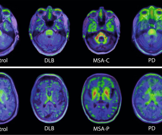
Swiss biopharmaceutical company AC Immune highlighted a study showing the potential of its alpha-synuclein (a-syn) PET tracer for diagnosing neurodegenerative disease. b) Transversal images at the level of the basal ganglia in a control participant, and patients with DLB, MSA-parkinsonian type. (b)
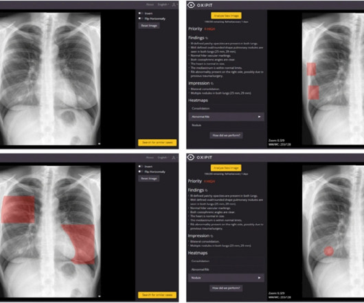
A study conducted in a primary care clinic in Spain highlights the need for improvement of AI algorithms for interpreting chest x-rays, despite being commercially approved by European regulators, according to an article published March 3 in Scientific Reports. Image courtesy of Scientific Reports. The full article is available here.
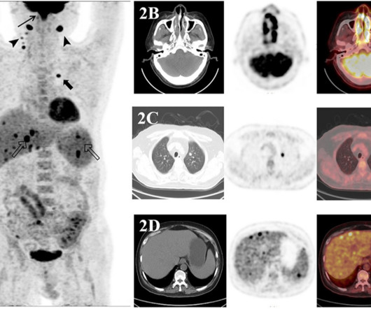
In a retrospective analysis, a team in Taiyuan, China, found that F-18 FDG-PET/CT was superior for analyzing imaging manifestations of NK/T-cell lymphoma and suggested that the scans can improve treatment plans for patients. “F-18 Transaxial images show an F-18 FDG-avid mass in both ethmoid sinus (B).
On July 9, 2024, AuntMinnieEurope.com posted an article about the extensive damage caused to Ukraine's largest pediatric hospital. Radiologist Josh Sokol, MD, a founder of the FRI who works with teleradiology company vRad, read AuntMinnieEurope.com 's July 9 article and recognized the urgent need for ultrasound capabilities at the Ohmatdyt.
CBBCT in recent years has garnered interest among radiologists as another way to diagnose breast cancer. The modality provides isotropic 3D images, which the researchers highlighted eliminates the need for breast compression. CBBCT uses a cone-shaped x-ray beam to acquire multiple 2D images from various angles around the breast.
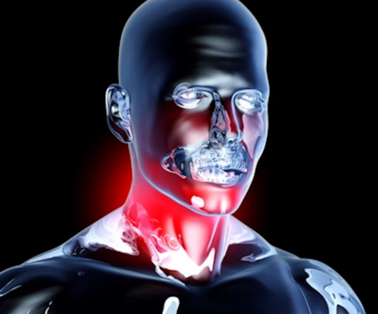
In an analysis of cases where F-18 FDG radiotracer uptake was reported as suspicious for oropharynx cancer on PET/CT scans, the number of patients who were actually diagnosed was low, the group found. Further analysis revealed that these diagnosed patients had higher F-18 FDG mean ipsilateral radiotracer uptake.
Strategies to increase the use of cardiac PET myocardial perfusion imaging (MPI) in the U.S. Cardiac PET MPI has emerged as a key tool for diagnosing and managing patients with cardiovascular diseases, especially coronary artery disease, yet overall it remains underutilized in the U.S., The full article can be found here.
PET/CT imaging can serve as a metabolic guide to increase the accuracy of needle biopsies in patients with suspicious lung nodules, according to a study published January 18 in the European Journal of Radiology. Image courtesy of the European Journal of Radiology. The full article can be found here. Greece, and the U.S.
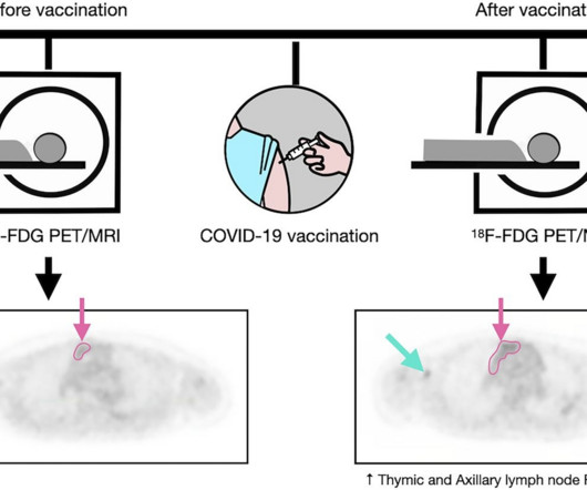
Understanding postvaccination imaging findings in patients with cancer is important because these results may be confounded with tumor relapse or metastasis,” wrote first author Gaurav Luthria, MD, PhD, and colleagues. Image courtesy of the Journal of Nuclear Medicine. The full article is available here.
A radiomics nomogram based on F-18 FDG-PET/CT imaging could help clinicians assess long-term outcomes in women undergoing treatment for locally advanced cervical cancer, according to a study published January 30 in BMC Cancer. The full article can be found here.
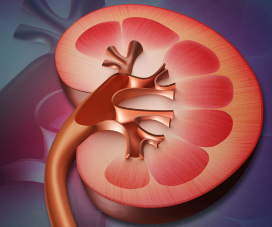
PSMA-PET/CT has the potential to be an effective alternative imaging approach for diagnosing patients with renal cell carcinoma (RCC), a type of kidney cancer, according to a study published May 23 in the Journal of Nuclear Medicine. Using a comprehensive search strategy, the authors initially identified 145 articles.
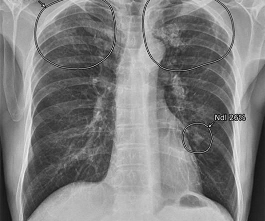
A group at Yonsei University College of Medicine in Gyeonggi-do, South Korea, evaluated how often clinically significant lung nodules were detected unexpectedly on chest x-rays by AI (Insight CXR, v3, Lunit ) and whether coexisting findings can aid in differential diagnoses. Image courtesy of Scientific Reports.
OpenAI's GPT-4 AI model can utilize imaging reports to generate summaries of disease course in patients with complex glioblastoma, improving treatment planning and potentially even enhancing radiology workflows, according to research published July 23 in Radiology. No image data were transmitted to GPT-4.
PET imaging using a newly developed radiotracer has identified different patterns of brain tau pathology over time in early-onset versus late-onset Alzheimer’s disease patients, according to a study published February 1 in the Journal of Nuclear Medicine. Image courtesy of the Journal of Nuclear Medicine. in 17 early-onset patients.
Endovascular thrombectomy (EVT) is effective for stroke patients who need to be transferred from hospitals where they are diagnosed to centers capable of performing the procedure, according to a study published February 8 in JAMA Network Open. The full article is available here. Out of 352 enrolled patients (median age, 66.5
Before we get further, I published a series of articles grouped here as a “ Guide to the Core Exam ” that lays out a lot of helpful information. We are radiologists, and the majority of the exam is image identification: you need to look at lots and lots of pictures.
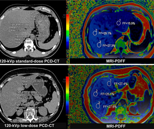
Photon-counting CT may serve as an alternative to MRI for assessing liver fat in patients with fatty liver disease, according to an article published September 24 in Radiology. A) Representative axial unenhanced 120-kVp standard-dose CT image (left) and corresponding MRI scan (right) in a 33-year-old man (body mass index, 26.2)
In clinical care, amyloid PET scans are used to diagnose and manage Alzheimer’s disease patients based on the amounts of beta-amyloid plaque the scans reveal in the brain. In previous evidence from the Imaging Dementia–Evidence for Amyloid Scanning (IDEAS) study, amyloid PET was associated with changes in patient management in 60.2%
A group at Mount Sinai Hospital developed a “pipeline” of convolutional neural networks (CNNs) to analyze lung areas in DDR image sequences from patients. To that end, the group developed two convolutional neural networks (CNN) designed to quantify key measurements in DDR image sequences. Image and caption courtesy of Chest Pulmonary.
A pair of articles published in the May issue of the Journal of Nuclear Medicine illustrate the promise of the novel FAPI radiotracer in diagnosing, staging, and treating multiple types of cancer.
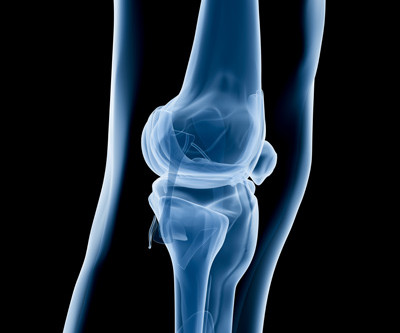
While MRI-detected biomarkers may serve a beneficial role for people with hip and knee pain, the modality's clinical utility may be limited in patients over the age of 45 in patients with advanced osteoarthritis, according to an article published in the American Journal of Roentgenology ( AJR ).
To that end, the researchers tested GPT-4’s ability to model the demographic diversity of medical diagnoses by constructing 10 unique prompts, each of which asked GPT-4 to generate an example patient presentation with a specific medical condition. The full article can be found here. prevalence estimates. based prevalence estimates.
Cureus is calling for submissions of research articles focusing o. Residents measure up when diagnosing pneumothorax Imaging, labs aid in treating teen with rare jaw tumor Imaging leads to removal of 3-cm toy from dementia patient Echo helps identify flossing-induced heart disease
UCLA researchers have developed a deep-learning framework that teaches itself quickly to automatically analyze and diagnose MRIs and other 3D medical images—with accuracy matching that of medical specialists in a fraction of the time.
New articles will be published each Monday until our official anniversary at RSNA 2024. Our top article in 2018 covered an important early development that supported efforts to train AI algorithms. Such algorithms can help teach computers how to detect and diagnose disease, the NIH said in a September 27 announcement.
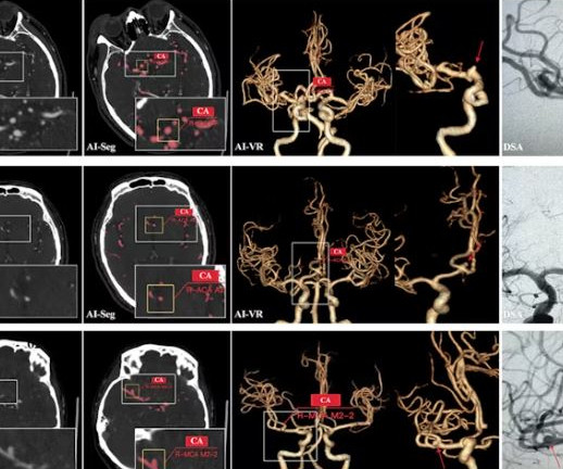
What’s more, the model performed consistently on images generated by different scanner manufacturers. However, cerebral aneurysm identification on CTA images is time-consuming and error-prone, particularly for less-experienced clinicians, due to the diminutive size and faint contrast of the aneurysm against cerebral vessels.
Risk factors Michael Alosco, PhD, of Boston University and the Chobanian and Avedisian School of Medicine is among those contributing to brain imaging and white matter research. Gray matter-white matter integrity Researchers used diffusion-tensor imaging, an MRI technique, to study the impact of soccer heading on the brain.
Over the past 100 years, medical imaging has become vital to almost every medical practice. Radiological equipment such as the X-ray machine provides a low-risk way for healthcare providers to deliver fast and accurate diagnoses for treatment guidance.
milla1cf Fri, 03/29/2024 - 08:01 March 29, 2024 — Magnetic resonance imaging ( MRI ) is a cornerstone in the landscape of medical diagnostics, celebrated for its non-ionizing and non-invasive nature. For more information: [link] Friday, March 29, 2024 - 08:01
lead author of the article and neuroradiologist at Mayo Clinic in Jacksonville, Florida. Increased use of monoclonal antibodies led to the discovery of amyloid-related imaging abnormalities (ARIA). Patients with ARIA sometimes have headaches, but they are usually asymptomatic and only diagnosable with MRI. “It Agarwal, M.B.B.S.,
Depending on your imaging protocols, perhaps that means you will open up the accompanying CT of the cervical spine, because the thinner slices on that exam may make a fracture more apparent even though it’s the brain that I’m reading right now. You can then individually save generated flashcards into an Anki deck.
The results offer additional incentives for using bpMRI to diagnose the disease, wrote a team led by Emmanuel Salinas-Miranda, MD, of the Ottawa Hospital in Canada. Access the full article here. The group's findings were published March 5 in the American Journal of Roentgenology.
The model performed well, and you can read about the study in this issue’s featured article. performed well on image-independent American College of Radiology Diagnostic In-Training Exam (ACR DXIT) practice questions. As expected, AI studies took most of the top headlines from RSNA 2023.
milla1cf Tue, 01/16/2024 - 14:37 January 16, 2024 — The Society of Nuclear Medicine and Molecular Imaging ( SNMMI ) and the European Association of Nuclear Medicine (EANM) have issued a new procedure standard/practice guideline for estrogen receptor imaging of breast cancer patients using FES PET.
We organize all of the trending information in your field so you don't have to. Join 5,000 users and stay up to date on the latest articles your peers are reading.
You know about us, now we want to get to know you!
Let's personalize your content
Let's get even more personalized
We recognize your account from another site in our network, please click 'Send Email' below to continue with verifying your account and setting a password.


Let's personalize your content