This site uses cookies to improve your experience. To help us insure we adhere to various privacy regulations, please select your country/region of residence. If you do not select a country, we will assume you are from the United States. Select your Cookie Settings or view our Privacy Policy and Terms of Use.
Cookie Settings
Cookies and similar technologies are used on this website for proper function of the website, for tracking performance analytics and for marketing purposes. We and some of our third-party providers may use cookie data for various purposes. Please review the cookie settings below and choose your preference.
Used for the proper function of the website
Used for monitoring website traffic and interactions
Cookie Settings
Cookies and similar technologies are used on this website for proper function of the website, for tracking performance analytics and for marketing purposes. We and some of our third-party providers may use cookie data for various purposes. Please review the cookie settings below and choose your preference.
Strictly Necessary: Used for the proper function of the website
Performance/Analytics: Used for monitoring website traffic and interactions
Our study showed that the availability of PET/CT prior to the [percutaneous needle lung biopsy] improves the diagnostic biopsy rates,” wrote lead author Konstantinos Stefanidis, MD, of King’s College Hospital in London, and colleagues. Patient with extensive lung and pleural disease. (2a) The full article can be found here.
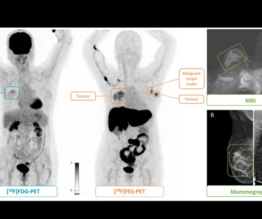
F-18 fluoroestradiol (FES) PET may improve staging of grade 1 or 2 estrogen receptor (ER)-positive breast cancer compared with F-18 FDG-PET, according to an article published March 4 in Radiology. Nuclear medicine physicians analyzed both sets of images and determined disease stages, with final stages verified via biopsy. (B)
RadiologyInfo.org , a source of medical imaging information for the general public, has introduced a new series of articles and videos to help patients understand their radiology exam reports. Those interested in the new series of articles and videos can visit the website.
“Skin biopsy is the gold standard for diagnosing calciphylaxis, but it can worsen lesions and confer poorer disease prognosis,” the researchers wrote, in a study published October 23 in JAAD International. The team assessed the potential diagnostic utility of bone scans in calciphylaxis based on a review of the literature.
The finding comes despite guidelines recommending LSG as the diagnostic test of choice and underlines the need for a better diagnostic test, wrote lead author Tina Moon, MD, of Tufts Medical Center in Boston and colleagues. The disease is a particular concern among cancer patients, they noted.
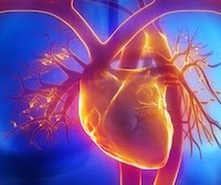
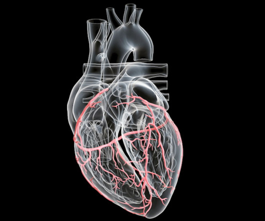
Cardiac PET MPI has emerged as a key tool for diagnosing and managing patients with cardiovascular diseases, especially coronary artery disease, yet overall it remains underutilized in the U.S., The full article can be found here. Strategies to increase the use of cardiac PET myocardial perfusion imaging (MPI) in the U.S.
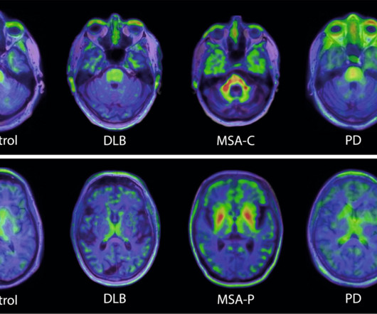
Swiss biopharmaceutical company AC Immune highlighted a study showing the potential of its alpha-synuclein (a-syn) PET tracer for diagnosing neurodegenerative disease. The finding indicates that the tracer could enable earlier, more accurate diagnosis of MSA and potentially, more precise monitoring of disease progression, AC Immune said.
We hope you enjoy our article as well as the opportunity to hear directly from the recipients. He earned his medical degree at the University of Alabama School of Medicine and completed a residency in diagnostic radiology at the University of Florida.
newspaper coverage, news articles over the past 25 years have discussed AI’s advantages more than potential risks. However, radiologists were “infrequently” interviewed or quoted in these articles. Radiologists continue to investigate and debate the potential of AI to improve diagnostic accuracy and clinical workflows.
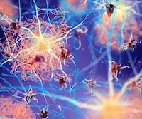
PET imaging has revealed brain pathology linked to faster clinical progression of Alzheimer’s disease in patients with ApoE4 gene variants, according to a study published November 6 in JAMA Neurology. A link to the full article can be found here. The study authors included scientists at Eli Lilly and Company.
According to the findings, there were significant differences in GPT-4's modeling of disease prevalence by race and gender compared with true U.S. Ultimately, there are real, biologically meaningful relationships between diseases and patient demographics, the researchers noted. The full article can be found here.
This incidental finding highlights the potential of AI-based decision support for patient management in terms of diagnostic and treatment planning based on amino acid PET,” noted lead author Philipp Lohmann, PhD, of Aachen University in Aachen, and colleagues. A link to the full case report can be found here.
The document was published jointly March 11 in the Journal of Nuclear Cardiology , Clinical Infectious Diseases , the Heart Rhythm Journal , and JACC: Cardiovascular Imaging. In the article, experts provide evidence-based consensus on specific clinical scenarios where PET/CT and SPECT/CT add value for patient care. In the U.S.
His research interests include using structural and functional MRI -- particularly ultrahigh-field, 7-tesla MRI -- to map brain microstructure and develop neurosurgical treatment of brain tumors, epilepsy, and neurodegenerative and movement disorders such as Parkinson's disease, essential tremor, and dystonia. He served in the U.S.
The low detection rate of primary tumors by current diagnostic techniques remains a major concern for patients with head and neck cancer of unknown primary,” the group wrote. Primary tumors were detected in 46 (51%) patients after a thorough diagnostic work-up. The full article is available here.
FDG-PET imaging shows promise for use as a diagnostic criterion for neurosarcoidosis, with a recent case series illustrating the approach was effective when gold-standard approaches were not, according to a group of researchers in Berlin. “To The article was published January 25 in Neurological Research and Practice.
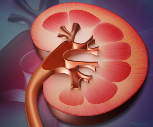
In a meta-analysis of published studies, a group at Johns Hopkins University School of Medicine in Baltimore, MD, found that PET/CT using both gallium-68 (Ga-68)-based and F-18-based prostate-specific membrane antigen (PSMA) radiotracers had high detection rates for staging primary RCC lesions and restaging metastatic or recurrent disease.
To make the indications more specific to and improve the overall diagnostic performance, F-18 FDG-PET/MRI should be indicated in FUO and IOU especially in patients with elevated CRP,” the authors noted. The full article is available here. Fever of unknown origin (FUO) is defined as a temperature higher than 38.3 °C
New articles will be published each Monday until our official anniversary at RSNA 2024. Our most highly read article in 2021 reported on two of these studies. Ultrasound from diagnostic work-up performed seven days later showed no change in lymph node size. BI-RADS category 0 was assigned. BI-RADS 3 was assigned.
Common diagnostic tests for pulmonary disorders include chest x-rays and pulmonary function tests (PFTs). The full article is available here. “Our findings add to growing evidence suggesting DDR as a potential [pulmonary function test] surrogate,” noted lead author and internal medicine fellow Valeria Santibanez, MD, and colleagues.
Thanks to its better noise reduction, DLR also maintains diagnostic image quality, according to the authors. We're seeing now is that cardiac CT is a fantastic test to rule out the presence of flow-limiting coronary disease," Leiner said, adding that the future is not about single algorithms.
0866T -- qMRI analysis of the brain with comparison to prior MR study(ies), including lesion detection, characterization, and quantification, with brain volume(s) quantification and/or severity score, when performed, data preparation and transmission, interpretation, and report, obtained with diagnostic MRI examination of the brain.
Eliot Siegel, MD; Stanislav Spiridonov, MD; Nathan Gee, MD; and Anthony Chang, PhD, are among a niche gathering of early adopters, entrepreneurial physicians, medical physicists, and investors with a sweet spot for nuclear medicine, diagnostic radiology, and radiation oncology.
What’s the difference between Screening and Diagnostic Mammogram? Screening mammography is a routine exam for women who have no sign or symptoms of breast disease. During a diagnostic mammogram, the images are analyzed in real-time. Additionally, the billing is different between screening and diagnostic mammograms.
asked Michael Morris, MD, of Memorial Sloan Kettering Cancer Center in New York for an article in the Journal of Nuclear Medicine (JNM) last year. How many centers have the physical space in their nuclear medicine departments to treat a disease as common as prostate cancer?" Morris continued in the JNM.
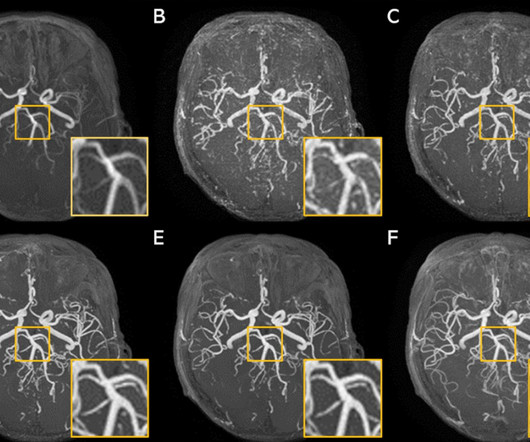
This study aimed to develop a generative adversarial network (GAN) model to improve the image resolution of brain time-of-flight MR angiography (TOF-MRA), as well as evaluate the image quality and diagnostic utility of the reconstructed images.
We hope you enjoy our article as well as the opportunity to hear directly from the recipients. Our hope is that we'll be able to determine how to use this as a diagnostic tool in actual practice,” he said. He added that by the age of 50 or 60, many or most people have already had a CT scan for diagnostic purposes. “We
milla1cf Fri, 03/29/2024 - 08:01 March 29, 2024 — Magnetic resonance imaging ( MRI ) is a cornerstone in the landscape of medical diagnostics, celebrated for its non-ionizing and non-invasive nature.
New articles will be published each Monday until our official anniversary at RSNA 2024. Our top article in 2017 covered an EU regulatory body's controversial recommendation to pull four gadolinium-based contrast agents from the market.
This retrospective study investigated whether volumetric visceral adipose tissue (VAT) features that were extracted using radiomics and three-dimensional convolutional neural network (3D-CNN) approaches are effective when differentiating Crohn’s disease (CD) and ulcerative colitis (UC).
When it comes to drilling in diagnostic radiology, I also interpret this to mean honing your search pattern/approach. And having models for different patterns of disease allows you to pick up on subtle constellations of findings. Drill Drill: Practice skills repeatedly until they become automatic and natural.
He has applied this investigative approach across a range of neurological disorders, with a major focus on stroke prevention and cerebrovascular disease assessment. Gupta has been recognized with numerous awards including the Distinguished Investigator Award from the Academy for Radiology & Biomedical Imaging Research and the Robert C.
The American College of Radiology (ACR) has tracked the movement of this national coverage determination (NCD) as previously reported and has advocated for removing existing barriers and providing broader access to beta-amyloid PET imaging for Alzheimer's Disease (AD) diagnosis, management and evaluation of newly approved therapies.
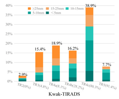
Dear Editor, We read with interest the article by Sun Huh et al [1] in the Jul 2021 issue of European Radiology. disease) in the enrolled sample is known to influence diagnostic tests [2]. Moreover, the threshold has been used to overcome the overdiagnosis of thyroid cancer, rather than enhance the diagnostic performance of RSS.
Teleradiology Introduction: Radiant smiles are not just the result of good oral hygiene; they are also a testament to the role of dental X-rays in supporting preventive and diagnostic dentistry. Gum Health Assessment: Periodontal disease often starts beneath the gumline. This aids in diagnosing hidden sources of pain or discomfort.
MRI-Scan-Teleradiology Introduction: Echocardiography, a non-invasive imaging technique, is the unsung hero of diagnostic cardiology. In this article, we will explore the indispensable role of echocardiography in diagnostic cardiology. It goes beyond the heart’s chambers, offering crucial insights into cardiac health.
The model performed well, and you can read about the study in this issue’s featured article. performed well on image-independent American College of Radiology Diagnostic In-Training Exam (ACR DXIT) practice questions. As expected, AI studies took most of the top headlines from RSNA 2023.
Teleradiology in a flat world Introduction: Cone Beam Computed Tomography (CBCT) technology has transcended its role in dentistry, extending its reach into the broader realm of medical diagnostics. Enhanced Dental Diagnostics: In dentistry, CBCT has revolutionized diagnostics.
Introduction: Austria, a country celebrated for its cultural heritage and scenic beauty, is taking significant strides in advancing diagnostics through the integration of teleradiology into its healthcare system. Teleradiology is revolutionizing diagnostic services, advancing Austrian medicine to new heights.
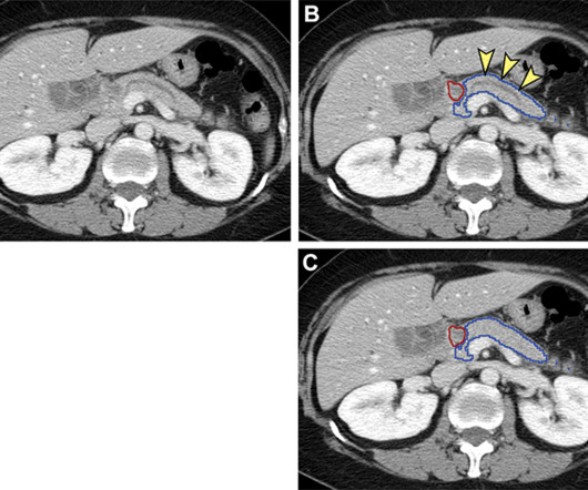
Margulis Award for Scientific Excellence will be presented to authors of the Radiology article, “ Pancreatic Cancer Detection on CT Scans with Deep Learning: A Nationwide Population-based Study.” Named for Alexander R. Margulis , M.D., We are truly honored and genuinely surprised to have received this award,” Dr. Chen said.
Benefits of Teleradiology to Telehealth Introduction: Australia, a land known for its diverse landscapes and healthcare challenges, is undergoing a significant transformation in diagnostics through the remarkable strides in teleradiology. Traditional radiology services often faced limitations, leading to delayed diagnoses and treatments.
Background: The increased utility and accessibility of point-of-care ultrasound (POCUS) has allowed clinicians the freedom to rethink their diagnostic approach for many common diseases, including peritonsillar abscess (PTA). Article: Kim DJ et al. First, Do No Harm! If inconclusive, order a CT scan.
Introduction: Armenia, a country known for its rich history and culture, is currently experiencing a remarkable diagnostic transformation through the invaluable contribution of teleradiology. Skilled Healthcare Workforce Armenia’s diagnostic transformation is driven by a highly skilled and dedicated healthcare workforce.
Introduction: Australia, known for its vast and diverse landscapes, is paving the way for the future of diagnostic imaging through the innovative use of teleradiology. Skilled Healthcare Workforce The future of diagnostic imaging in Australia is driven by a highly skilled and dedicated healthcare workforce.
We organize all of the trending information in your field so you don't have to. Join 5,000 users and stay up to date on the latest articles your peers are reading.
You know about us, now we want to get to know you!
Let's personalize your content
Let's get even more personalized
We recognize your account from another site in our network, please click 'Send Email' below to continue with verifying your account and setting a password.


Let's personalize your content