This site uses cookies to improve your experience. To help us insure we adhere to various privacy regulations, please select your country/region of residence. If you do not select a country, we will assume you are from the United States. Select your Cookie Settings or view our Privacy Policy and Terms of Use.
Cookie Settings
Cookies and similar technologies are used on this website for proper function of the website, for tracking performance analytics and for marketing purposes. We and some of our third-party providers may use cookie data for various purposes. Please review the cookie settings below and choose your preference.
Used for the proper function of the website
Used for monitoring website traffic and interactions
Cookie Settings
Cookies and similar technologies are used on this website for proper function of the website, for tracking performance analytics and for marketing purposes. We and some of our third-party providers may use cookie data for various purposes. Please review the cookie settings below and choose your preference.
Strictly Necessary: Used for the proper function of the website
Performance/Analytics: Used for monitoring website traffic and interactions
We hope you enjoy our article as well as the opportunity to hear directly from the recipients. Importantly, we are gaining deeper insights into disease processes themselves, which enhances our ability to diagnose and potentially treat conditions that were previously beyond our reach." I'm a radiographer,' " Stewart recalled.
a) Raw example of a dynamic digital radiograph. (b) Moreover, while PFTs are vital for diagnosing and monitoring pulmonary disorders, they pose challenges in accessibility, such as in patients with neuromuscular conditions or those experiencing flares of chronic obstructive pulmonary disease, they suggested.
This algorithm represents the first step to automatically classify and organize shoulder radiographs on a large scale in very little time, which will profoundly enrich shoulder arthroplasty registries,” the group wrote, in an article published October 23 in Journal of Shoulder and Elbow Surgery.
His research interests include using structural and functional MRI -- particularly ultrahigh-field, 7-tesla MRI -- to map brain microstructure and develop neurosurgical treatment of brain tumors, epilepsy, and neurodegenerative and movement disorders such as Parkinson's disease, essential tremor, and dystonia.
Alzheimer disease is a progressive, irreversible brain disorder that slowly degrades memory and cognitive function. While previous treatment methods focused on addressing Alzheimer disease symptoms, recent approvals of monoclonal antibodies have provided a path to target the underlying disease itself. In June 2021, the U.S.
The Society publishes five established journals: Radiology, RadioGraphics, Radiology: Artificial Intelligence, Radiology: Cardiothoracic Imaging and Radiology: Imaging Cancer. RadioGraphics , RSNA’s premier educational journal, edited by Christine ‘Cooky’ Menias , M.D., Klein , M.D., RSNA Board Liaison for Publications.
We hope you enjoy our article as well as the opportunity to hear directly from the recipients. Of course, no awards ceremony is complete without acceptance speeches. So this year we’ve invited our victors to say a few words about their Minnies wins. We’re starting to see the fruits of these goals and it’s good,” Menias said.
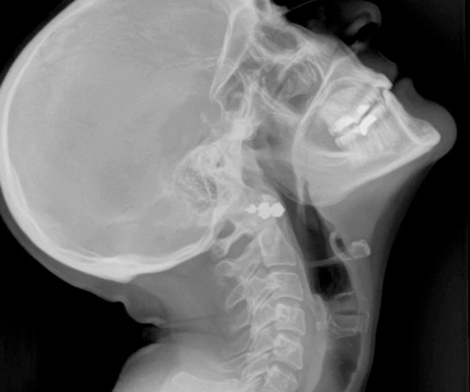
The article titled “Craniofacial Manifestations of Systemic Disorders: CT and MR Imaging Findings and Imaging Approach” discusses imaging approaches for early detection of various systemic diseases or conditions that affect the maxillofacial bones.
The article titled “Craniofacial Manifestations of Systemic Disorders: CT and MR Imaging Findings and Imaging Approach” discusses imaging approaches for early detection of various systemic diseases or conditions that affect the maxillofacial bones.
DDR could also provide a more comprehensive understanding of a patient’s neuromuscular component of ventilatory disorders and many other pulmonary diseases.” I'm particularly interested in seeing if it will be helpful for patients with certain neuromuscular diseases such as myasthenia gravis.
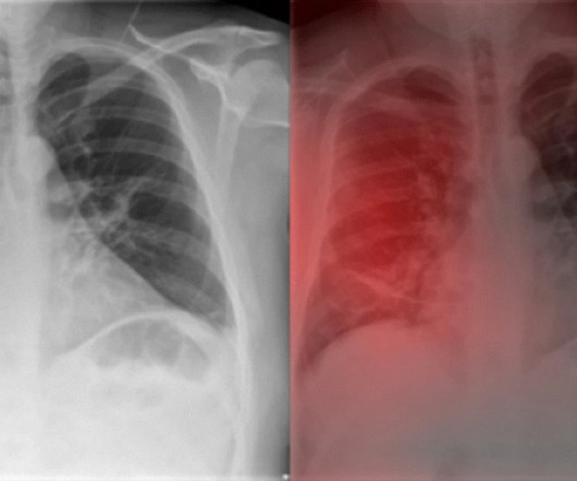
However, the sensitivity of a chest radiograph in the diagnosis of COVID-19 is moderate. The authors were able to show that the performance of Ensemble4Covid from the onset of the disease was considerably higher compared to the radiologists.
Article: Marx T, Joly LM, Parmentier AL, et al. Thoracentesis with free drainage for 15 minutes Drainage at -25cm H20 for 30 minutes Clamping and radiograph; If failed, 2nd aspiration at -25cm H20 for 30 minutes, Repeat clamping and radiograph. Am J Respir Crit Care Med. 2023;207(11):1475-1485. PMID: 20696690 Baumann, M.
Rose The prevalence of osteoarthritis (OA) in dogs is nearly 40%, and radiographic evidence of the condition is found in 90% of cats aged 12 or older. Read More Diagnostic Imaging Systems does not claim any ownership or take any credit for the articles listed in the Veterinary E-News Magazine. Read More Lame Horse?
Article: de-Madaria E et al. Primary Safety Endpoint: Fluid Overload defined by 2 of the following 3: Criterion 1: non-invasive evidence of heart failure (ie echo), radiographic evidence of pulmonary congestion, invasive cardiac Cath suggesting heart failure. Baseline disease severity was well-balanced between groups.
Centers for Disease Control and Prevention (CDC) is considering the use of AI to back up its tuberculosis (TB) screening program for immigrants and refugees. After COVID-19, TB is the second leading cause of death from infectious disease in the world. The full article can be found here. has relatively low rates – about 2.5
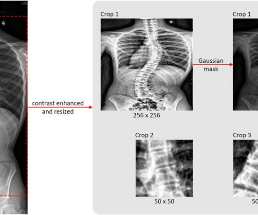
Treatment options depend on the severity of disease and range from observation alone to bracing and surgery. Three regions of interest (ROI) were selected and processed for training from each original radiograph. The leftmost image is the original coronal view radiograph. The full article is available here.
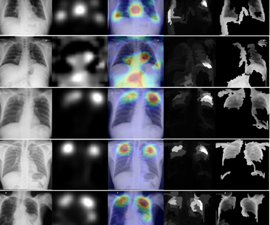
accurate labeling x-ray reports for thoracic diseases, but performed poorly in patients over 80 years old, noted lead author Samantha Santomartino, a medical student at Drexel University in Philadelphia, and colleagues. Three board-certified radiologists annotated the images for 14 thoracic disease labels.
They are crucial for detecting various diseases such as cancer. In some cases, early detection of other diseases can potentially save a life. This article will talk about the different diagnostic imaging methods such as X-rays, CT scans, Ultrasound, and MRI. X-rays are also used to diagnose infections, tumors, and bone disease.
Photoprint from radiograph by W.K. 3) In the early twentieth century, it was a common goal for investigators to try to find a way to separate the superimposed shadows that were recorded when a complex structure was shown on a radiograph. (3) This is now known as ‘Hand mit Ringen’. (1) after seeing the image. (2) Röntgen, 1895.
Article : Costa O.S. POPULATION Inclusions: Age ≥ 18 years of age Radiographically confirmed acute spontaneous or traumatic intracranial hemorrhage (i.e. Given the natural history of the disease process, patients may have had similar outcomes without treatment. The primary outcome —hemostatic effectiveness— is disease-oriented.
Diagnosis and Management of Cerebral Small Vessel Disease. 0000000000001232 Cerebral small vessel disease (CSVD) is one of the most common clinical conditions that a neuroimager will encounter. It has well-defined radiographic features and various clinical presentations. Anti-amyloid antibody therapies in Alzheimer’s disease.
The goal of this article is to discuss the indications and limitations as well as to provide a basic guide to interpretation of noncontrast CT imaging of the brain (NCCT), CT angiography (CTA) of the head and neck, and CT perfusion (CTP) imaging in acute stroke evaluation. Vascular Diseases of the Brain.” Radiographics.
We organize all of the trending information in your field so you don't have to. Join 5,000 users and stay up to date on the latest articles your peers are reading.
You know about us, now we want to get to know you!
Let's personalize your content
Let's get even more personalized
We recognize your account from another site in our network, please click 'Send Email' below to continue with verifying your account and setting a password.


Let's personalize your content