This site uses cookies to improve your experience. To help us insure we adhere to various privacy regulations, please select your country/region of residence. If you do not select a country, we will assume you are from the United States. Select your Cookie Settings or view our Privacy Policy and Terms of Use.
Cookie Settings
Cookies and similar technologies are used on this website for proper function of the website, for tracking performance analytics and for marketing purposes. We and some of our third-party providers may use cookie data for various purposes. Please review the cookie settings below and choose your preference.
Used for the proper function of the website
Used for monitoring website traffic and interactions
Cookie Settings
Cookies and similar technologies are used on this website for proper function of the website, for tracking performance analytics and for marketing purposes. We and some of our third-party providers may use cookie data for various purposes. Please review the cookie settings below and choose your preference.
Strictly Necessary: Used for the proper function of the website
Performance/Analytics: Used for monitoring website traffic and interactions
New articles will be published each Monday until our official anniversary at RSNA 2024. MRI safety articles have always been popular with our members, so it’s not surprising that our top article in 2019 reported on a safety incident in Sweden. Following the incident, metal detector gates were installed, he said.
We hope you enjoy our article as well as the opportunity to hear directly from the recipients. He is particularly excited about the growth of ultrahigh field MRI in neuroimaging -- especially since he reads neurodegenerative and movement disorder cases every day, he told AuntMinnie.com. "We I'm a radiographer,' " Stewart recalled.
His research interests include using structural and functional MRI -- particularly ultrahigh-field, 7-tesla MRI -- to map brain microstructure and develop neurosurgical treatment of brain tumors, epilepsy, and neurodegenerative and movement disorders such as Parkinson's disease, essential tremor, and dystonia.
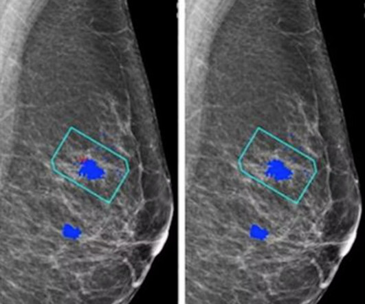
For the AI study, two views of each breast (four images) are taken to be evaluated independently either by the AI system plus a single reader or by a radiologist/radiographer as a first reader and second reader using the two-reader model. when using an AI system to differentiate cancers from benign lesions at breast MRI.
The full article is available here. The study highlights “the importance of how a one-week radiology boot camp can impact both future radiology and non-radiology clerkship experiences,” the group concluded.
We hope you enjoy our article as well as the opportunity to hear directly from the recipients. The gatekeeper with risk will look at MRI and CT and ask, 'Does that study really matter to the care decision?' Of course, no awards ceremony is complete without acceptance speeches. And they're going to chip away at waste."
lead author of the article and neuroradiologist at Mayo Clinic in Jacksonville, Florida. Patients with ARIA sometimes have headaches, but they are usually asymptomatic and only diagnosable with MRI. “It Most patients with asymptomatic ARIA meeting specific radiographic and clinical criteria may continue to receive treatment.
Available exclusively online, the journal will offer continuous publication, featuring primarily original multidisciplinary research articles with a focus on emerging topics, cross-cutting and innovative research. She has served on the RSNA Genitourinary Scientific Program Committee and RadioGraphics Genitourinary Review Panel.
Kim Mason Kim Mason, an Audit and Research Radiographer for Mid Yorkshire Teaching Hospitals Trust, talks about their role as well as the value of radiographer engagement in research activities and how to get involved. Hi, I’m Kim and I am an alternative-styled, funky-haired, septum-pierced, disabled Audit and Research Radiographer.
MRI-Scan-Teleradiology Introduction: Dental radiography is an essential component of modern dentistry, offering valuable insights into oral health and guiding treatments with precision. Quality Assurance: Dental radiographers are meticulous in quality assurance. Let’s explore the key aspects of this intricate process.
In these women, screening tests, such as ultrasound or MRI, when added to mammography, substantially increase the detection of early-stage breast cancer. A growing number of medical organisations link to the DenseBreast-info.org website, including the EFRS (European Federation of Radiographer Societies) and the Society of Radiographers.
I’ve published articles on the topic. Preamble – a personal perspective I’ve chaired committees tasked with writing MCQs for high-stakes radiology exams. I’ve taught the art of writing good MCQs both locally and nationally. I’ve personally written hundreds of items.
Rather, we may see a staged move from AI-based coreading (serving as a secondary check, especially for high-volume or after-hours work) to more autonomous reading -- starting in settings such as overnight or emergency radiology before expanding into daytime general practice and, eventually, subspecialty domains like complex MRI. Meta AI Blog.
This article will talk about the different diagnostic imaging methods such as X-rays, CT scans, Ultrasound, and MRI. X-ray Also called a radiograph, an X-ray uses radiation to create images of the body. When getting an MRI, the patient needs to remain very still for the scanner to produce high-quality images.
Photoprint from radiograph by W.K. 3) In the early twentieth century, it was a common goal for investigators to try to find a way to separate the superimposed shadows that were recorded when a complex structure was shown on a radiograph. (3) Their work gave rise to the modern MRI scanners we use today. after seeing the image. (2)
It has well-defined radiographic features and various clinical presentations. They described six radiographic phenotypes of CSVD: (1) recent small subcortical infarct, (2) white matter hyperintensity, (3) lacune of presumed vascular origin, (4) widened perivascular spaces, (5) cerebral microbleed, and (6) brain atrophy. 2023;2015(5).
The goal of this article is to discuss the indications and limitations as well as to provide a basic guide to interpretation of noncontrast CT imaging of the brain (NCCT), CT angiography (CTA) of the head and neck, and CT perfusion (CTP) imaging in acute stroke evaluation. Radiographics. Last Update: March 22 2019.
We organize all of the trending information in your field so you don't have to. Join 5,000 users and stay up to date on the latest articles your peers are reading.
You know about us, now we want to get to know you!
Let's personalize your content
Let's get even more personalized
We recognize your account from another site in our network, please click 'Send Email' below to continue with verifying your account and setting a password.


Let's personalize your content