This site uses cookies to improve your experience. To help us insure we adhere to various privacy regulations, please select your country/region of residence. If you do not select a country, we will assume you are from the United States. Select your Cookie Settings or view our Privacy Policy and Terms of Use.
Cookie Settings
Cookies and similar technologies are used on this website for proper function of the website, for tracking performance analytics and for marketing purposes. We and some of our third-party providers may use cookie data for various purposes. Please review the cookie settings below and choose your preference.
Used for the proper function of the website
Used for monitoring website traffic and interactions
Cookie Settings
Cookies and similar technologies are used on this website for proper function of the website, for tracking performance analytics and for marketing purposes. We and some of our third-party providers may use cookie data for various purposes. Please review the cookie settings below and choose your preference.
Strictly Necessary: Used for the proper function of the website
Performance/Analytics: Used for monitoring website traffic and interactions
More than 60% of diagnostic radiology and radiation therapy staff experience workplace violence, according to a study published January 9 in Radiography. To address this knowledge gap, the authors reviewed medical literature to identify articles on the issue over the last 10 years. The full article is available here.
AuntMinnie.com recently spoke with Warren Gefter, MD, of the University of Pennsylvania, lead author of a recent article in Radiology proposing that human-AI symbiosis – rather than totally autonomous AI – should be the current goal for AI in chest radiography.
Researchers at the Icahn School of Medicine at Mount Sinai ("Icahn Mount Sinai") used Dynamic Digital Radiography (DDR) data, an X-ray imaging technology developed by Konica Minolta, to create their AI-powered technique that analyzes lung function. Chest radiography is typically acquired in the evaluation of pulmonary disorders.
The article was published March 7 in IEEE Transactions on Medical Imaging. However, the general concepts behind the corrections presented here are applicable for all grating-based radiography systems, including even the recently presented clinical dark-field CT system,” the researchers concluded.
In addition, they computed the out-of-network rate by imaging modality, with claims categorized as CT, MR, nuclear medicine (NM), ultrasound, radiography or fluoroscopy (XR), and interventional procedures. The full article can be found here. In total, they identified 80.4 million imaging claims for 19.5
Before we get further, I published a series of articles grouped here as a “ Guide to the Core Exam ” that lays out a lot of helpful information. And, of course, the question of how to pass the Core Exam after a failure is mostly the same as asking how to pass it in the first place.
Music can be calming for patients undergoing medical imaging, an article published February 7 in Radiography suggests. This includes imaging procedures such as MRI, mammography, and PET among others.
Chest dynamic digital radiography (DDR) may have received a boost toward clinical use in patients with lung disorders, with researchers developing AI to perform time-consuming analysis involved in the technology, according to researchers in New York City. The full article is available here.
He has published more than 1,000 articles, is the author or co-author of 10 textbooks, and member of editorial boards for more than 35 journals. She is a registered technologist in radiography and received her radiologic technology degree from Drexel University in Philadelphia.
The new discovery lit a fire in the scientific community, and was so sensational that in the following year over 1,000 articles would be published on the topic of X-rays. Radiography has exploded into a variety of modalities and specialisms from CT to Ultrasound to MRI; all driven by research and development. How can I get involved?
Since the cortical bone can be seen on panoramic radiography, this area of the lower jaw canal may be helpful in assessing BMD. In this cross-sectional study, black pixel intensity on the radiography was used to measure BMD of the mandibular canal cortices.
Introduction : In the past decade, veterinary medicine has witnessed a transformative shift with the adoption of digital radiography systems in place of traditional film-based methods. For veterinary professionals contemplating the switch to digital radiography, this article offers essential guidance to ensure a seamless transition.
MRI-Scan-Teleradiology Introduction: Dental radiography is an essential component of modern dentistry, offering valuable insights into oral health and guiding treatments with precision. Patient-Centered Approach: Dental radiography starts with the patient. Continuing Education: Staying current with the latest advances is paramount.
The model performed well, and you can read about the study in this issue’s featured article. An AI algorithm was presented that could make dynamic digital radiography (DDR) more efficient by automatically measuring kinematics involved in certain shoulder injuries. As expected, AI studies took most of the top headlines from RSNA 2023.
We hope you enjoy our article as well as the opportunity to hear directly from the recipients. UNC-Chapel Hill developed collaborative agreements with UNC Health and Duke Health for the planning and execution of two programs, a certificate program in radiography, and a bachelor of science degree program in diagnostic medical sonography.
Project philosophical doughnut was an editorial project to tidy up, standardize and, most importantly, give clinical indications and purpose to our CT protocol articles (hence the wacky name Andrew chose and stuck with).
In this article, we’ll take a journey through time to explore how technology has revolutionized the world of dental X-rays, providing more precision, efficiency, and safety for both patients and dental professionals. Nowhere is this more evident than in the evolution of dental X-ray equipment.
About the author Vincent Chan is President and General Manager of Digital Radiography at Carestream Health. A version of this article previously appeared in DOTmed magazine. Ultimately, participating in conversations around collaboration and innovation can help the industry achieve these shared goals.
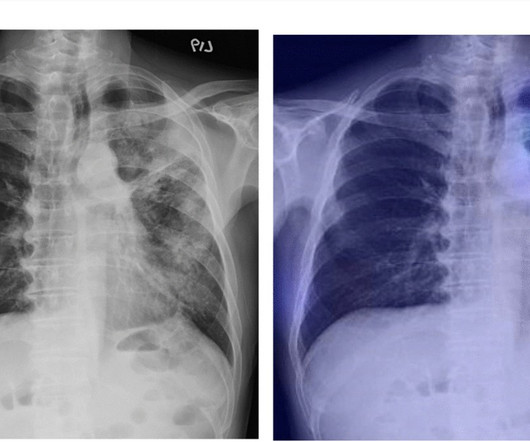
This was especially seen in the role of chest radiography when it was utilized as a diagnostic tool at the beginning of the pandemic “when microbiological resources were scarce,” evolving into its use focusing on the detection and monitoring of COVID-19 lung involvement.
In this article, we will explore the latest innovations that are shaping the future of dental X-ray imaging, revolutionizing patient care and clinical practices. Digital Radiography: Traditional film-based X-ray imaging is making way for digital radiography.
This technology has paved the way for navigating the third dimension, offering a transformative role in imaging that transcends the limitations of conventional two-dimensional radiography. In this article, we will embark on a journey through the fascinating world of CBCT and its impact on the field of diagnostic imaging.
Article: A deep learning model using chest X-ray for identifying TB and NTM-LD patients: a cross-sectional study Authors: Chia-Jung Liu, Cheng Che Tsai, Lu-Cheng Kuo, Po-Chih Kuo, Meng-Rui Lee, Jann-Yuan Wang, Jen-Chung Ko, Jin-Yuan Shih, Hao-Chien Wang & Chong-Jen Yu DNN model could be a screening tool for mycobacterial lung diseases.
Article: The diagnostic performance and clinical value of deep learning-based nodule detection system concerning influence of location of pulmonary nodule Authors: Seulgi You, Ji Hyun Park, Bumhee Park, Han-Bit Shin, Taeyang Ha, Jae Sung Yun, Kyoung Joo Park, Yongjun Jung, You Na Kim, Minji Kim & Joo Sung Sun
The unfortunate result is that most mammographers were and are not currently learning techniques and principles that apply to positioning of all other radiography exams. 1) Look for those who have published articles in respected journals. 1) Look for DATA and/or articles that prove their techniques produce better mammograms ( 3 )!
Article: Marx T, Joly LM, Parmentier AL, et al. Absence of residual pneumothorax or the presence of an apical residual pneumothorax smaller than 2 cm, based on chest radiography. Simple Aspiration versus Drainage for Complete Pneumothorax: A Randomized Noninferiority Trial. Am J Respir Crit Care Med. 2023;207(11):1475-1485.
Although a retrospective study can eliminate some bias of image acquisition as discussed above, prospective studies tend to give us better information on a topic and there have been several which refute this article’s conclusions. One of the largest, Chan et. Acad Emerg Med. 2005 Sep;12(9):844-9. doi: 10.1197/j.aem.2005.05.005.
About the Author Vincent Chan is President and General Manager of Digital Radiography at Carestream. A version of this article was originally published on AuntMinnie.com The post 3 AI-Based Technology Trends in Medical Imaging appeared first on Everything Rad.
As a Professor of Practice (Diagnostic and Interventional Radiology) since 2016, at the National Heart & Lung Institute, Imperial College London, he maintains an active academic career, publishing widely with over 220 articles in peer reviewed journals.
It is important to emphasize that the EUSOBI recommendations highlighted in this article are not yet guidelines in Europe. EUSOBI acknowledges that it may take time before the new recommendations are implemented in Europe and that the level of implementation is dependent on the resources that are available locally.
According to the article, there is currently no ultrasound scanner or x-ray machine at the Obongi Health Centre IV, the main health facility in Obongi District in Uganda, and this is having a major impact on both the local population and the 130,000 refugees from South Sudan who are currently also served by the facility.
“These findings contribute to a better understanding of the radiation dosimetry associated with different patient positions during WB-PET/CT scans,” the group wrote, in an article published January 4 in Radiography.
This article is dedicated to comparing these rules and providing a reasonable guide for maximizing their individual utility. The provided infographics detail the specifics of each rule for quick reference. Ann Emerg Med. 2013 May;61(5):521-7. Mower WR, Gupta M, Rodriguez R, Hendey GW. 2017 Jul 11;14(7):e1002313. Pediatr Radiol.
We have simply gathered top stories from some of the best veterinary news sources to share with our customers, along with updated veterinary conference information, and special equipment promotions.
We organize all of the trending information in your field so you don't have to. Join 5,000 users and stay up to date on the latest articles your peers are reading.
You know about us, now we want to get to know you!
Let's personalize your content
Let's get even more personalized
We recognize your account from another site in our network, please click 'Send Email' below to continue with verifying your account and setting a password.


Let's personalize your content