This site uses cookies to improve your experience. To help us insure we adhere to various privacy regulations, please select your country/region of residence. If you do not select a country, we will assume you are from the United States. Select your Cookie Settings or view our Privacy Policy and Terms of Use.
Cookie Settings
Cookies and similar technologies are used on this website for proper function of the website, for tracking performance analytics and for marketing purposes. We and some of our third-party providers may use cookie data for various purposes. Please review the cookie settings below and choose your preference.
Used for the proper function of the website
Used for monitoring website traffic and interactions
Cookie Settings
Cookies and similar technologies are used on this website for proper function of the website, for tracking performance analytics and for marketing purposes. We and some of our third-party providers may use cookie data for various purposes. Please review the cookie settings below and choose your preference.
Strictly Necessary: Used for the proper function of the website
Performance/Analytics: Used for monitoring website traffic and interactions
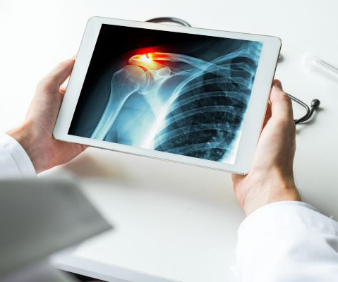
Bones become more fragile and porous with age, so less force is needed to break their structure. Medical imaging is used to help diagnose these injuries, so doctors can propose appropriate treatment plans. While X-rays are typically utilized, an MRI or CT scan may be recommended. What Are Broken Bones?
Imagine going in for a diagnostic MRIscan and coming out with a progressive, irreversible, and potentially fatal disease. Last week, attorneys for a Cleveland man announced they had filed suit against a gadolinium manufacturer, claiming that he developed NSF from an injection of gadolinium MRI contrast. 356: 9234, pp.
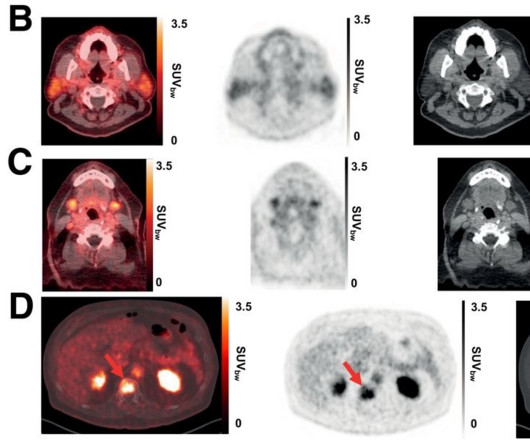
for imaging PSMA-positive lesions in patients with newly diagnosed or recurrent prostate cancer, yet the biodistribution of these tracers can vary, the authors explained. Overall, more participants with potential tumor lesions and more potential lesions were detected with Ga-68 PSMA-R2 PET/CT than with conventional imaging, the group wrote.
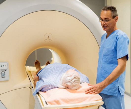
Hospitals and medical practices of all sizes, no matter their target demographic or geographical location, often struggle to ensure their patients have access to all of the essential imaging modalities necessary to diagnose a wide variety of medical conditions. The answer to many of these questions could be just one—MRI.
As I was on the phone with a colleague trying to convince the referrer of why I think a patient has Paget’s disease instead of metastases, I described the cortical thickening of the iliopectineal line and the lack of activity on the bonescan at the site and elsewhere throughout the body.
MRI is key to diagnosis, obtain this imaging in all patients who raise clinical suspicion Patients with hemodynamic instability and neurologic compromise warrant empiric antibiotics. Imaging Gadolinium enhanced MRI – modality of choice, highly sensitive and specific ( Mylona 2009 ).
Prostate cancer persists as the most frequently diagnosed malignancy in men beyond skin cancer. In this new editorial, researchers Richard L.J. Qiu, Chih-Wei Chang, and Xiaofeng Yang from Emory University discuss prostate cancer. To design the salvage radiation therapy, imaging is required to detect and locate the recurrence disease regime.”
Nuclear medicine is a form of specialty medicine that uses radioactive tracers to evaluate bodily functions and to diagnose and treat a wide range of health conditions. Nuclear scans produce images of the body’s anatomy that cannot be obtained as clearly or fully with other imaging techniques.
Doctors use imaging tests to see inside a patient’s body and diagnose their illnesses and injuries. There are several types of imaging tests that physicians use to detect cancer in patients: X-Ray, Computed Tomography (CT), Magnetic Resonance Imaging (MRI), Ultrasound (US), Nuclear Medicine, and Positron Emission Tomography (PET).
Bonescan shows diffuse uptake in the markedly enlarged right breast secondary to increased blood pool activity and impaired washout. Bonescan showed diffuse uptake in the markedly enlarged right breast secondary to increased blood pool activity and impaired washout. Mammogram CC view. Surg Clin North Am. Breast Cancer.
A bone density test is a non-invasive procedure to measure the strength of your bones. It is an effective way to diagnosebone-related health problems, such as osteoporosis, and to assess the risk of developing them. DEXA Bone Density Scan A bone density scan is carried out using a DEXA bone densitometry machine.
We organize all of the trending information in your field so you don't have to. Join 5,000 users and stay up to date on the latest articles your peers are reading.
You know about us, now we want to get to know you!
Let's personalize your content
Let's get even more personalized
We recognize your account from another site in our network, please click 'Send Email' below to continue with verifying your account and setting a password.


Let's personalize your content