This site uses cookies to improve your experience. To help us insure we adhere to various privacy regulations, please select your country/region of residence. If you do not select a country, we will assume you are from the United States. Select your Cookie Settings or view our Privacy Policy and Terms of Use.
Cookie Settings
Cookies and similar technologies are used on this website for proper function of the website, for tracking performance analytics and for marketing purposes. We and some of our third-party providers may use cookie data for various purposes. Please review the cookie settings below and choose your preference.
Used for the proper function of the website
Used for monitoring website traffic and interactions
Cookie Settings
Cookies and similar technologies are used on this website for proper function of the website, for tracking performance analytics and for marketing purposes. We and some of our third-party providers may use cookie data for various purposes. Please review the cookie settings below and choose your preference.
Strictly Necessary: Used for the proper function of the website
Performance/Analytics: Used for monitoring website traffic and interactions
The new horizons of advanced imaging, especially in oncology, must increasingly consider not only diagnostic efficacy but also sustainability for patients, prize-winning researchers told ECR 2025 delegates. To mitigate these effects, low-energy imaging techniques should be prioritized without compromising diagnostic accuracy.
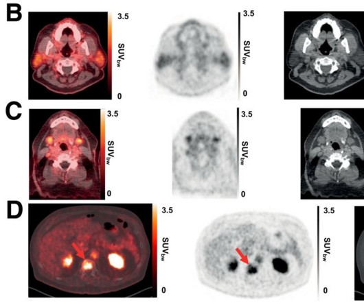
In the trial, 30 patients with recurrent disease or metastatic prostate cancer underwent baseline conventional imaging (CT, MRI, or bonescan) and whole-body Ga-68 PSMA-R2 PET/CT between 20 minutes and four hours after injection. All patients had undergone either radical prostatectomy or radiation therapy. (A)
HeartLungs AutoBMD AI opportunistically detects osteoporosis in CT scans done for other reasons so the patient receives no extra X-ray radiation or scanning time. Osteoporosis screening guidelines updated: Do you need a bonescan? Bone Health and Osteoporosis Foundation. References: 1. Sudhakar S.
Imagine going in for a diagnostic MRI scan and coming out with a progressive, irreversible, and potentially fatal disease. Diagnostic and Interventional Radiology , December 2006, Vol. Anteroposterior and lateral Tc-99m HDP delayed bonescan shows extensive symmetric skin uptake in lower extremities and distal upper extremities.
Although the patient is exposed to radiation during the procedure, the test is considered safe because the amount of radiation used is comparable to that of a routine X-ray. This scan may be performed in order to check whether you have a suspected kidney obstruction or impaired function.
Our Women's Center performs both screening and diagnostic mammograms. Ultrasound is a simple, safe, painless diagnostic procedure that bounces high-frequency sound waves off parts of the body and captures the returning “echoes” as images. There is no injection or radiation exposure associated with ultrasound.
There are no special instructions to prepare for it, and unlike an MRI or CT Scan , a bone densitometry procedure does not involve lying in an enclosed tunnel. The scan uses a very low dose of radiation to produce pictures of the inside of the body, particularly around the hips and lower spine, to measure bone mass and strength.
We organize all of the trending information in your field so you don't have to. Join 5,000 users and stay up to date on the latest articles your peers are reading.
You know about us, now we want to get to know you!
Let's personalize your content
Let's get even more personalized
We recognize your account from another site in our network, please click 'Send Email' below to continue with verifying your account and setting a password.


Let's personalize your content