This site uses cookies to improve your experience. To help us insure we adhere to various privacy regulations, please select your country/region of residence. If you do not select a country, we will assume you are from the United States. Select your Cookie Settings or view our Privacy Policy and Terms of Use.
Cookie Settings
Cookies and similar technologies are used on this website for proper function of the website, for tracking performance analytics and for marketing purposes. We and some of our third-party providers may use cookie data for various purposes. Please review the cookie settings below and choose your preference.
Used for the proper function of the website
Used for monitoring website traffic and interactions
Cookie Settings
Cookies and similar technologies are used on this website for proper function of the website, for tracking performance analytics and for marketing purposes. We and some of our third-party providers may use cookie data for various purposes. Please review the cookie settings below and choose your preference.
Strictly Necessary: Used for the proper function of the website
Performance/Analytics: Used for monitoring website traffic and interactions
Technetium-99m (Tc-99m) methyl diphosphonate (MDP) bonescans are a potentially viable noninvasive option for diagnosing calciphylaxis, according to a team at the University of Massachusetts in Worcester, MA. The team assessed the potential diagnostic utility of bonescans in calciphylaxis based on a review of the literature.
"Whole-body MRI is emerging as a superior tool for assessing bone marrow and monitoring therapy response in advanced breast cancer. It can detect progressive disease in cases missed by other modalities, demonstrating its potential to guide treatment decisions effectively," they noted. DL2 had the shortest mean scan time (0.27 0.04
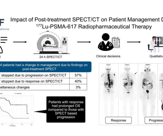
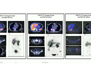
Yet whether SPECT/CT can be used to determine disease progression or response has yet to be delineated, they noted. To that end, the group assessed the impact of SPECT/CT imaging on 122 patients who had received a minimum of two cycles of Pluvicto and had post-treatment scans after each cycle.
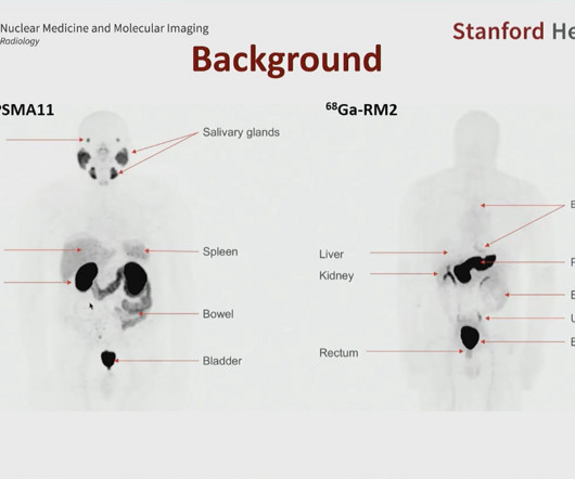
Moreover, the approach could help detect disease that other effective prostate cancer PET radiotracers may miss, said Heying Duan, MD, a nuclear medicine research scientist at Stanford University, during a November 26 scientific session. Patients were eligible if they were older than 18 and had rising blood levels of PSA ( ≥ 0.2
The primary outcome was the correct identification of the prostate cancer tumor stage, which is crucial for surgeons to determine the extent of the disease -- whether it has spread beyond the prostate gland, for instance. Out of 150 men who participated, 134 ultimately underwent radical prostatectomies (the mean age was 62 years old).
PET/CT imaging with a new gallium-68-based prostate cancer radiotracer shows promise for detecting recurrent metastatic disease, according to a study published February 20 in the Journal of Nuclear Medicine. The finding is from a phase I/II clinical trial in the U.S.
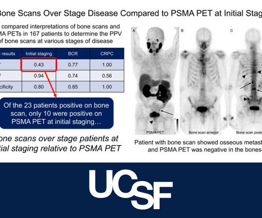
An article in the Journal of Nuclear Medicine reports findings of a multi-center retrospective study that compares bonescan with PSMA PET for initial staging of prostate cancer.
As I was on the phone with a colleague trying to convince the referrer of why I think a patient has Paget’s disease instead of metastases, I described the cortical thickening of the iliopectineal line and the lack of activity on the bonescan at the site and elsewhere throughout the body.
To design the salvage radiation therapy, imaging is required to detect and locate the recurrence disease regime.” However, the introduction of 18F-fluciclovine (anti-1-amino-3-18F-fluorocyclobutane-1-carboxylic acid) PET/CT has marked a significant advancement in salvage disease management.
The degree of elevation does not predict disease severity. CRP concentration rise and fall quicker than ESR, often used to guide treatment Blood Cultures – an important element in management and treatment Blood culture positivity often decides whether a patient will require a bone biopsy. ESR and CRP are sensitive, yet not specific.
3-Phase BoneScan A 3-phase bonescan is used to diagnose abnormal areas within the bones or joints, such as a fracture, bone pain, infection, or bonedisease, which cannot be seen successfully with a standard X-ray.
Bonescan shows diffuse uptake in the markedly enlarged right breast secondary to increased blood pool activity and impaired washout. Bonescan showed diffuse uptake in the markedly enlarged right breast secondary to increased blood pool activity and impaired washout. Mammogram CC view.
Capitol Imaging Services offers a variety of ultrasound imaging tests, like: Liver Elastography Ultrasound Guided Breast Biopsy Nuclear Medicine Nuclear Medicine imaging scans utilize a small, harmless dose of a radioactive isotope to highlight parts of the body, making them easier to see in an imaging test.
Osteoporosis Osteoporosis is a disorder which affects the bones and is characterized by a progressive loss of bone tissue. This weakens bones making them porous, fragile, and susceptible to fractures. Osteoporosis is a common bonedisease with as many as 1.5
Although studies have shown that PSMA-PET is highly effective for detecting prostate cancer, the long-term consequences of widely implementing the technique in patients with recurrent disease are unknown, the authors explained. population and was informed by data from real-world settings.
In addition, the mean tissue dose did not reach the maximum tolerated dose for kidney, bone marrow, or liver. Three grade 3 CAM-H2 treatment emergent adverse events (TEAE) were reported in three patients who had a previous history of thrombocytopenia, lung disease, or liver disease, with no grade 4 or 5 adverse events reported.
Imagine going in for a diagnostic MRI scan and coming out with a progressive, irreversible, and potentially fatal disease. It certainly doesn't help matters that the disease has been linked to gadolinium-based contrast agents, a ubiquitous tool in the MR armamentarium. In 2006 the U.S. Louis hospital.
Even if an X-ray shows a fracture, your doctor may recommend a CT scan or MRI for better visualization of damaged tissue and other secondary injuries. BoneScans A bonescan is a type of nuclear imaging test that allows for better observation of certain bonediseases.
The findings suggest a better alternative to "conventional" imaging modalities such as CT alone, MRI, or bonescans for tracking prostate cancer in this particular patient population, wrote a team led by Adrien Holzgreve, MD, of the University of California, Los Angeles. The results were published January 3 in JAMA Network Open. "[Our
We organize all of the trending information in your field so you don't have to. Join 5,000 users and stay up to date on the latest articles your peers are reading.
You know about us, now we want to get to know you!
Let's personalize your content
Let's get even more personalized
We recognize your account from another site in our network, please click 'Send Email' below to continue with verifying your account and setting a password.


Let's personalize your content