This site uses cookies to improve your experience. To help us insure we adhere to various privacy regulations, please select your country/region of residence. If you do not select a country, we will assume you are from the United States. Select your Cookie Settings or view our Privacy Policy and Terms of Use.
Cookie Settings
Cookies and similar technologies are used on this website for proper function of the website, for tracking performance analytics and for marketing purposes. We and some of our third-party providers may use cookie data for various purposes. Please review the cookie settings below and choose your preference.
Used for the proper function of the website
Used for monitoring website traffic and interactions
Cookie Settings
Cookies and similar technologies are used on this website for proper function of the website, for tracking performance analytics and for marketing purposes. We and some of our third-party providers may use cookie data for various purposes. Please review the cookie settings below and choose your preference.
Strictly Necessary: Used for the proper function of the website
Performance/Analytics: Used for monitoring website traffic and interactions
The new horizons of advanced imaging, especially in oncology, must increasingly consider not only diagnostic efficacy but also sustainability for patients, prize-winning researchers told ECR 2025 delegates. To mitigate these effects, low-energy imaging techniques should be prioritized without compromising diagnostic accuracy.
mtaschetta-millane Tue, 07/02/2024 - 09:53 July 2, 2024 — A new editorial paper was published in Oncoscience ( Volume 11 ) on May 20, 2024, entitled, “ Deep learning-assisted lesion segmentation in PET/CT imaging: A feasibility study for salvage radiation therapy in prostate cancer.” In this new editorial, researchers Richard L.J.
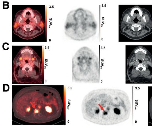
PET/CT imaging with a new gallium-68-based prostate cancer radiotracer shows promise for detecting recurrent metastatic disease, according to a study published February 20 in the Journal of Nuclear Medicine. Imaging agents with low uptake in at-risk organs and higher tumor uptake are needed, they noted.
At Clermont Radiology’s Women’s Center, we have created a soothing, spa-like environment staffed with caring experts, to meet all your Women’s Imaging needs. By listening closely to our patients, we have identified 3 “must-haves” that create an ideal women’s imaging experience: Comfort, Convenience, and Confidence.
Nuclear scans produce images of the body’s anatomy that cannot be obtained as clearly or fully with other imaging techniques. Nuclear medicine tests use a very small amount of radioactive tracer (radionuclide or radioisotope) which is specific for the organ or tissue to be scanned. We look forward to serving you.
The scan uses a very low dose of radiation to produce pictures of the inside of the body, particularly around the hips and lower spine, to measure bone mass and strength. A certified radiologic technologist will prepare your images for the radiologist, who will then interpret your results and prepare a report.
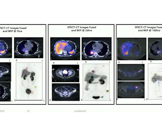
Theranostics is increasingly being used to image and treat prostate cancer and neuroendocrine tumors. Several presenters at the Society for Nuclear Medicine and Molecular Imaging's (SNMMI) 2024 meeting in Toronto shared new findings in women's studies. and included limited single and cumulative radiation doses.
How are imaging experts and others handling NSF? The FDA asked physicians to use alternative methods to image patients with moderate to end-stage renal disease whenever possible. Imaging is generally required for organs other than the kidneys and MR is the first-line imaging exam. Clearly, a major problem is at hand.
This AI solution will help Americans become aware of their bone loss before they face negative consequences such as bone fracture. "We HeartLungs AutoBMD AI opportunistically detects osteoporosis in CT scans done for other reasons so the patient receives no extra X-ray radiation or scanning time. References: 1.
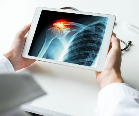
While broken bones are common injuries experienced across all ages, older individuals are at higher risk. Bones become more fragile and porous with age, so less force is needed to break their structure. Medical imaging is used to help diagnose these injuries, so doctors can propose appropriate treatment plans.
We organize all of the trending information in your field so you don't have to. Join 5,000 users and stay up to date on the latest articles your peers are reading.
You know about us, now we want to get to know you!
Let's personalize your content
Let's get even more personalized
We recognize your account from another site in our network, please click 'Send Email' below to continue with verifying your account and setting a password.


Let's personalize your content